Anxiolytic Effects of Onion Peel Quercetin, Aloe and Doum Palm Tannins Loaded Chitosan Nanoparticles in Carboxymethyl Cellulose-Induced Oxidative Stress in Albino Mice
-
Cletus Anes Ukwubile


Department of Pharmacognosy, Faculty of Pharmacy, University of Maiduguri, Maiduguri, Nigeria
Uwakmfon Idorenyin BasseyDepartment of Pharmacognosy, Faculty of Pharmacy, University of Maiduguri, Maiduguri, Nigeria
| Received 15 Apr, 2025 |
Accepted 16 Sep, 2025 |
Published 31 Dec, 2025 |
Background and Objective: Anxiety disorders, often linked to oxidative stress, are a major public health concern worldwide. While benzodiazepines are commonly prescribed, their use is limited by adverse effects such as sedation and dependency. This study aimed to evaluate the anxiolytic and antioxidant potential of onion peel-derived quercetin, Aloe vera extract and doum palm tannins encapsulated in chitosan nanoparticles (QATCNPs) using a carboxymethyl cellulose (CMC)-induced oxidative stress model in albino mice. Materials and Methods: QATCNPs were synthesized using the ionic gelation method and characterized by Fourier-transform infrared spectroscopy (FTIR), scanning electron microscopy (SEM) and dynamic light scattering (DLS), revealing a mean particle size of 150.32 nm. Thirty albino mice were divided into five groups (n = 6): Control, CMC-only, diazepam (2 mg/kg), chitosan NPs (50 mg/kg) and QATCNPs (50 mg/kg). Behavioral tests, including the elevated plus maze (EPM) and open field test (OFT), assessed anxiolytic behavior. Biochemical assays quantified oxidative stress markers: malondialdehyde (MDA), catalase (CAT), superoxide dismutase (SOD) and glutathione (GSH). Physiological and neurochemical markers were also measured. Data are presented as Mean±SE and statistical differences among groups were analyzed using One-Way ANOVA followed by Tukey’s post-hoc test (p<0.05). Results: The QATCNP-treated group spent significantly more time in the open arms of the EPM (150.00±5.00 sec) and center of the OFT (170.00±5.00 sec) compared to the CMC-only group (50.00±3.00 and 40.00±3.00 sec, respectively). The MDA levels decreased to 2.10±0.10 nmol/mg protein in the QATCNP group compared to 4.50±0.12 nmol/mg in the CMC group. Antioxidant enzyme levels increased significantly: CAT (32.00±1.20 U/mg), SOD (18.00±0.80 U/mg) and GSH (8.20±0.50 μmol/mg) in QATCNP-treated mice. Furthermore, GABA, serotonin and neuropeptide Y levels were significantly elevated in the QATCNP group, indicating neurochemical modulation. Conclusion: QATCNPs significantly reduced anxiety-like behavior and oxidative stress while enhancing neuroprotective markers, suggesting their potential as a safe and effective therapeutic strategy for anxiety management.
| Copyright © 2025 Ukwubile and Bassey. This is an open-access article distributed under the Creative Commons Attribution License, which permits unrestricted use, distribution, and reproduction in any medium, provided the original work is properly cited. |
INTRODUCTION
Anxiety disorders are among the most prevalent mental health conditions globally, impacting an estimated 284 million people across diverse populations and age groups1 These disorders encompass a spectrum of conditions, including generalized anxiety disorder, panic disorder, social anxiety disorder and specific phobias. Left untreated, anxiety disorders can lead to severe complications, including depression, substance abuse and even an increased risk of cardiovascular diseases2.
The underlying mechanisms driving anxiety disorders are multifactorial and involve complex interactions between genetic, environmental and neurobiological factors. Among these, oxidative stress has emerged as a critical contributor to the pathogenesis and progression of anxiety disorders3. Oxidative stress results from an imbalance between the overproduction of reactive oxygen species (ROS) and the body's ability to neutralize these reactive molecules through endogenous antioxidant defenses4. Elevated ROS levels cause cellular damage, including lipid peroxidation, protein oxidation and DNA fragmentation. Within the central nervous system (CNS), oxidative stress disrupts neuronal function, impairs neurotransmitter signaling and compromises neuroplasticity, all of which exacerbate anxiety symptoms5. Current pharmacological treatments for anxiety disorders primarily include benzodiazepines and selective serotonin reuptake inhibitors (SSRIs)6. While these medications are effective in symptom management, they are often associated with significant drawbacks. Benzodiazepines, for instance, provide rapid relief but can lead to sedation, cognitive impairment, dependency and withdrawal symptoms with prolonged use2. Consequently, the limitations of existing treatments necessitate the exploration of safer and more effective alternatives.
In recent years, natural products have gained considerable attention for their potential in managing anxiety disorders3. These compounds, derived from plants and other natural sources, often possess antioxidants, anti-inflammatory and neuroprotective properties that can alleviate anxiety symptoms. Flavonoids such as quercetin, polysaccharides from Aloe vera and tannins from doum palm are particularly noteworthy7. Quercetin a prominent flavonoid found in onion peels, quercetin exhibits potent antioxidant activity by scavenging free radicals, inhibiting lipid peroxidation and upregulating endogenous antioxidant enzymes such as superoxide dismuTBSe (SOD), catalase (CAT) and glutathione peroxidase (GPx)8. Aloe vera, known for its broad therapeutic properties, Aloe vera contains bioactive compounds that exhibit anti-inflammatory, immunomodulatory and antioxidant effects, which may contribute to its anxiolytic potential9. Doum palm tannins extracted from doum palm (Hyphaene thebaica), tannins have demonstrated neuroprotective effects, including the modulation of oxidative stress and neuroinflammation, which are central to the pathogenesis of anxiety disorders10.
Nanotechnology has emerged as a transformative approach to addressing the challenges associated with phytochemical delivery11. Among various nanocarriers, chitosan nanoparticles stand out due to their biocompatibility, biodegradability and ability to encapsulate bioactive compounds efficiently. Chitosan, a natural polysaccharide derived from chitin, offers several advantages, including enhanced solubility, stability and bioavailability of encapsulated compounds12. Moreover, chitosan nanoparticles enable controlled release and targeted delivery, ensuring the sustained and localized therapeutic action of the encapsulated phytochemicals13. This study aims to evaluate the anxiolytic and antioxidant effects of a novel formulation comprising quercetin, Aloe vera and doum palm tannins encapsulated in chitosan nanoparticles (QATCNPs).
MATERIALS AND METHODS
Study area: This study was carried out from October to December, 2024 at the research laboratory of the Department of Pharmacology and Toxicology, Faculty of Pharmacy, University of Maiduguri, Nigeria. The laboratory conditions were as stated in the guidelines for research laboratories by the University Regulatory Commission in Nigeria.
Materials: Chemicals and reagents: 99.1% purity chitosan powder of high molecular weight (ChemSaver, USA), sodium tripolyphosphate (TPP), quercetin (onion peel extract), Aloe vera gel, doum palm tannins, carboxymethyl cellulose (CMC) (Sigma Aldrich, USA) and ELISA kits (ThermoFisher Scientific, USA) for oxidative stress assay.
Synthesis of QATCNPs: The quercetin, Aloe vera and doum palm tannins-loaded chitosan nanoparticles (QATCNPs) were synthesized using the ionic gelation method14. Characterization of QATCNPs was performed using dynamic light scattering (DLS) for particle size and zeta potential, scanning electron microscopy (SEM) for surface morphology and Fourier-transform infrared spectroscopy (FTIR) to confirm the encapsulation of bioactive compounds14.
Experimental design: The study utilized a total of 30 albino mice, which were randomly divided into five groups, each consisting of six mice (n = 6 per group) as shown below:
| • | Group 1 (control group): The control group received normal saline (0.5 mL/day) orally throughout the experimental period | |
| • | Group 2 (CMC-only group): This group was treated with CMC (200 mg/kg orally), which served as the negative control | |
| • | Group 3 (standard drug group): Mice in this group were treated with diazepam (2 mg/kg, intraperitoneally), which served as the positive control | |
| • | Group 4 (chitosan nanoparticles group): This group was administered blank chitosan nanoparticles (50 mg/kg, orally) | |
| • | Group 5 (QATCNPs group): The mice in this group received QATCNPs (50 mg/kg, orally) |
All treatments were administered daily for three weeks under standard laboratory conditions. Behavioral tests, including the elevated plus maze (EPM) and open field test (OFT), were conducted to assess anxiolytic effects. At the end of the treatment period, the mice were euthanized and tissue samples were collected for biochemical analysis of oxidative stress markers such as malondialdehyde (MDA), catalase (CAT), superoxide dismuTBSe (SOD) and glutathione (GSH)1.
Behavioral assessments: To evaluate the anxiolytic effects of QATCNPs, two widely used behavioral assays, the elevated plus maze (EPM) and open field test (OFT), were conducted1.
Elevated plus maze (EPM): The EPM apparatus consisted of two open arms (exposed and unprotected) and two closed arms (enclosed by high walls) extending from a central platform, elevated 50 cm above the floor. Each mouse was placed individually in the center of the maze, facing one of the open arms and allowed to explore freely for 5 min. During this period, the time spent in the open arms versus the closed arms was recorded. Anxiety-like behavior was inferred from the proportion of time spent in the open arms. A lower duration in the open arms indicated higher anxiety levels, as rodents naturally avoid open, elevated spaces due to fear of predation. Conversely, increased exploration of the open arms was considered a sign of reduced anxiety15.
Open field test (OFT): The OFT was conducted in a square arena with marked grids and distinct zones, including a central area and peripheral regions. Each mouse was placed individually in the center of the arena and allowed to explore for 5 min. Key parameters recorded included locomotor activity (measured by the number of grid crossings) and the time spent in the central zone. Mice with higher anxiety levels tend to restrict their movement to the peripheral zones of the arena, avoiding the exposed central area. Reduced anxiety was reflected by increased time spent in the central zone and enhanced exploratory behavior. Locomotor activity was also evaluated to distinguish between anxiolytic effects and potential sedation1.
Biochemical analysis: Biochemical analysis was conducted to evaluate oxidative stress markers in the brain tissues of mice treated with QATCNPs and other experimental groups. The following procedures were performed for the analysis of malondialdehyde (MDA), catalase (CAT), superoxide dismuTBSe (SOD) and glutathione (GSH), utilizing spectrophotometric methods to ensure accuracy and reliability16.
Sample preparation: After behavioral assessments, mice were euthanized and their brains were quickly removed, washed with ice-cold phosphate-buffered saline (PBS) and weighed. The brain tissues were homogenized in 10% (w/v) ice-cold PBS using a tissue homogenizer. The homogenates were centrifuged at 10,000 rpm for 15 minutes at 4°C and the supernatants were collected for biochemical assays.
Malondialdehyde (MDA) assay: The MDA, a marker of lipid peroxidation, was measured using the Thiobarbituric acid reactive substances (TBARS) method16.
Catalase (CAT) activity assay: The CAT activity was measured by monitoring the decomposition of hydrogen peroxide (H2O2)17.
Superoxide dismuTBSe (SOD) activity assay: The SOD activity was determined using the nitro blue tetrazolium (NBT) reduction method. The inhibition of formazan formation by SOD was measured spectrophotometrically at 560 nm. Enzyme activity was expressed as units per mg of protein18.
Glutathione (GSH) assay: The GSH levels were quantified using the Ellman’s reagent method19.
Evaluation of physiological markers
Heart rate measurement: Heart rate was measured using a non-invasive tail-cuff method with a computerized plethysmograph20.
Blood Pressure Measurement: Blood pressure was also determined using the tail-cuff method. After acclimatization, systolic and diastolic blood pressures were recorded in a quiet environment to reduce stress-induced variability. The measurements were taken using a calibrated sensor that detects changes in blood flow as pressure is applied to the tail20.
Cortisol levels measurement: Cortisol levels were quantified using a commercially available Enzyme-Linked Immunosorbent Assay (ELISA) kit, following the manufacturer’s instructions21.
Evaluation of biochemical markers
Gamma-aminobutyric acid (GABA) levels: The GABA, the primary inhibitory neurotransmitter in the brain, was measured to evaluate the anxiolytic potential of the treatment. Brain tissue samples were homogenized in an ice-cold phosphate buffer (pH 7.4) and centrifuged at 12,000 rpm for 15 min at 4°C to obtain the supernatant. The GABA concentration was determined using a High-Performance Liquid Chromatography (HPLC) system with a UV detector. Pre-column derivatization with o-phthalaldehyde (OPA) enhanced the sensitivity of the GABA detection. Standard solutions of known GABA concentrations were used to create a calibration curve and the results were expressed in μg/mg of protein22.
Serotonin (5-HT) levels: Serotonin, a key neurotransmitter associated with mood regulation, was analyzed in the brain tissue to assess the treatment’s impact on serotonergic activity. Brain homogenates were prepared in ice-cold acidified buffer and centrifuged at high speed to isolate the supernatant. Serotonin levels were quantified using an Enzyme-Linked Immunosorbent Assay (ELISA) kit specific for 5-HT, following the manufacturer’s instructions23.
Neuropeptide Y (NPY) levels: NPY, a stress-responsive neuropeptide involved in emotional and behavioral regulation, was measured in brain homogenates. The samples were processed in a lysis buffer containing protease inhibitors, followed by centrifugation to separate the clear supernatant. NPY levels were determined using a sandwich ELISA kit24.
Statistical analysis: The data were expressed as the mean±SE. To compare the differences between multiple groups, One-way ANOVA followed by Tukey's post-hoc test was performed. The value of p<0.05 was taken as statistically significant.
RESULTS
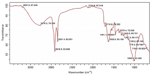
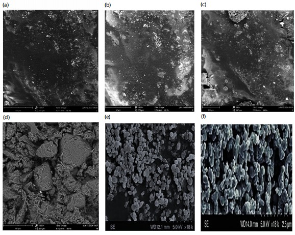
Nanoparticle characterization: The characterization of the formulated chitosan nanoparticles (QATCNPs) revealed promising properties for effective drug delivery applications. As shown in Table 1, QATCNPs 5 exhibited the highest percentage yield (56.66%) and entrapment efficiency (86.14%), indicating their superior encapsulation capacity compared to the other formulations. This suggests that QATCNPs 5 is the most efficient in retaining the bioactive components of the plant extract, likely due to the optimal chitosan-to-extract ratio and interaction. The FTIR analysis (Fig. 1) confirmed the successful encapsulation of phytochemicals into the chitosan matrix by identifying characteristic functional groups, such as hydroxyl, amine and carboxyl, which are indicative of both chitosan and plant-derived compounds. These functional groups may also contribute to the stability and bioactivity of the nanoparticles. Scanning electron microscopy (SEM) images (Fig. 2a-f) revealed that all formulations, including QATCNPs 5, had a spherical morphology with a smooth surface, indicating uniform particle formation and good dispersion characteristics. The regular morphology is crucial for predictable drug release and improved cellular uptake. Dynamic light scattering (DLS) analysis reported an average particle size of 150.24±2.14 nm for QATCNPs 5, with a zeta potential of +25 mV. The nanoscale size favors cellular internalization, while positive zeta potential enhances stability by preventing particle aggregation due to electrostatic repulsion. QATCNPs 5 also demonstrated an impressive cumulative drug release of 98.44% over 24 hrs, following first-order kinetics, which implies a concentration-dependent release ideal for sustained therapeutic action. The release profile was optimal under acidic conditions (pH 4.2), which mimics the tumor microenvironment or inflamed tissues, making QATCNPs 5 particularly suitable for targeted drug delivery in such conditions.
Behavioral outcomes: In the EPM test (Table 2), the QATCNP-treated group showed a significant increase in the time spent in the open arms of the maze compared to the CMC-only group (p<0.05). This result indicates reduced anxiety-like behavior, as the mice in the QATCNP group exhibited a greater willingness to explore the open, unprotected arms, a behavior typically avoided by anxious mice. In contrast, the CMC-only group, which was subjected to oxidative stress, spent more time in the closed arms, reflecting higher anxiety levels. The positive control group (diazepam-treated) also showed increased time in the open arms, confirming the anxiolytic effect of the standard treatment.
Similarly, in the OFT test (Table 2), the QATCNP-treated group demonstrated a significantly higher amount of time spent in the central area of the arena compared to the CMC-only group (p<0.05). Mice with reduced anxiety levels typically explore the central zone, which is considered a less safe area, more than anxious mice that tend to stay in the peripheral regions. The CMC-only group spent significantly less time in the center, reflecting the anxiety induced by oxidative stress. Similar results were observed in the diazepam-treated group, indicating that the QATCNP treatment had a comparable effect to the standard anxiolytic drug in reducing anxiety-like behavior.
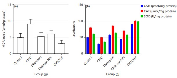
Effects on oxidative stress markers: Figure 3a below shows the MDA levels in the brain tissue. The results revealed that the QATCNP group demonstrates a significant (p<0.05) reduction in MDA levels compared to the CMC-only group, highlighting the antioxidative effect of the treatment. In Fig. 3b, the activities of CAT, SOD and GSH were compared across the different groups. The results show that the QATCNP group exhibits significantly higher activities of these antioxidant enzymes compared to the CMC-only group, indicating enhanced antioxidant defense.
|
|
Effects on physiological and biochemical markers: The results in Table 3 show that the QATCNP-treated group showed a reduced heart rate compared to the CMC-only group, indicating improved cardiovascular health. Also, the systolic blood pressure (mmHg) reveals that QATCNP treatment significantly lowered systolic pressure, approaching control levels, while the diastolic blood pressure (mmHg) also showed similar improvements for the QATCNP-treated group. Reduced cortisol levels in the QATCNP-treated group reflect lower stress was also noticed (Table 3). Similarly, in the biochemical markers (Table 4), QATCNPs enhanced GABA levels significantly compared to the CMC-only group, demonstrating anxiolytic potential. There were also elevated serotonin levels in the QATCNP-treated group, suggesting improved mood regulation as well as increased NPY levels in the QATCNP group, indicating better stress resilience (Table 4).
|
| Table 1: | Characterization of formulated chitosan NPs (QATCNPs) | |||
| Characterization parameter | Observation | Explanation |
| Yield (EE (%)) | QATCNPs 1: 24.12 (30.11) QATCNPs 2: 26.04 (44.18) QATCNPs 3: 14.12 (38.12) QATCNPs 4: 34.01 (42.04) QATCNPs 5: 56.66 (86.14) QATCNPs 6: 44.08 (52.08) |
QATCNPs 5 has the highest percentage yield regarding initial weights of chitosan plus plant extracts |
| FTIR analysis | Functional groups confirmed successful phytochemical encapsulation |
FTIR spectrum indicated the presence of key functional groups, confirming the successful encapsulation of phytochemicals into nanoparticles |
| SEM morphology a smooth surface |
Spherical nanoparticles with have a spherical shape and a smooth surface, indicating uniformity in morphology |
SEM images revealed that the nanoparticles |
| DLS size | Average particle size: 150.24±2.14 nm, Zeta potential: +25 mV |
Particle size distribution measured by DLS showed an average size of 150.24 nm, with a narrow range (±10 nm) and the positive zeta potential (+25 mV) indicates good stability of the nanoparticles in suspension |
| Cumulative drug release in 24 hrs (for QATCNPs 5) |
98.44% | Maintained sustained release following first-order kinetics |
| pH | 4.2 | More drugs were released under acidic conditions by QATCNPs 5 |
| SEM: Scanning electron microscope, EE (%): Percentage entrapment efficiency, FTIR: Fourier transform infrared spectroscopy, DLS: Dynamic light scattering and QATCNPs 1-6 are various chitosan NPs formulations | ||
| Table 2: | Behavioral outcomes in elevated plus maze (EPM) and open field test (OFT) | |||
| Treatment (group) | Time spent in open arms (EPM) (sec) | Time spent in center (OFT) (sec) |
| Normal control | 120.00±5.00 | 140.00±6.00 |
| CMC-only | 50.00±3.00 | 40.00±2.50 |
| Diazepam (2 mg/kg) | 130.00±4.00* | 135.00±5.00* |
| Chitosan NPs (50 mg/kg) | 110.00±4.50* | 120.00±4.00* |
| QATCNPs (50 mg/kg) | 150.00±5.00* | 170.00±6.00* |
| Values are expressed as Mean±SE, *Indicates statistical significance compared to the CMC-only group and determined by One-way ANOVA followed by Tukey’s post-hoc test (p<0.05) | ||
| Table 3: | Effects on physiological markers of the mouse brain tissue | |||
| Group | Heart rate (BPM) | Systolic BP (mmHg) | Diastolic BP (mmHg) | Cortisol levels (ng/mL) |
| Control | 350.00±2.02 | 110.00±1.11 | 70.00±0.04 | 3.50±0.01 |
| CMC-only | 420.00±2.01* | 140.00±1.03* | 90.00±0.14* | 5.00±0.01* |
| Diazepam (2 mg/kg) | 360.00±2.01 | 115.00±2.01 | 75.00±1.10 | 3.20±0.01 |
| Chitosan NPs (50 mg/kg) | 370.00±2.12 | 120.00±1.18 | 80.00±1.02 | 3.40±0.01 |
| QATCNPs (50 mg/kg) | 340.00±2.10* | 105.00±1.11* | 65.00±1.01* | 2.80±0.10* |
| *Indicates statistical significance at p<0.05 (One-way ANOVA followed by Turkey’s post hoc test) compared to the CMC-only group and Results are Mean±SD (n = 3) | ||||
| Table 4: | Effects on biochemical markers of the mouse brain tissue | |||
| Group | GABA levels (μg/mg protein) | Serotonin levels (ng/mg protein) | NPY levels (pg/mg protein) |
| Control | 15.00±1.01 | 50.00±1.03 | 100.00±2.04 |
| CMC-only | 8.00±0.01* | 30.00±1.01* | 60.00±1.11* |
| Diazepam (2 mg/kg) | 18.00±1.08 | 60.00±1.14 | 120.00±2.02 |
| Chitosan NPs (50 mg/kg) | 16.00±1.14 | 55.00±1.22 | 110.00±2.06 |
| QATCNPs (50 mg/kg) | 20.00±1.08* | 65.00±1.12* | 130.00±0.04* |
| *Indicates statistical significance at p<0.05 (One-way ANOVA followed by Turkey’s post hoc test) compared to the CMC-only group and Results are Mean±SD (n = 3) | |||
DISCUSSION
The findings of this study, which demonstrate the anxiolytic effects of QATCNPs as observed through behavioral improvements in the elevated plus maze (EPM) and open field test (OFT), align with previous studies that have highlighted the potential of individual phytochemicals, such as quercetin, Aloe vera and doum palm tannins, in alleviating anxiety. Previous research has shown that quercetin, a flavonoid with potent antioxidant properties, can reduce anxiety in rodent models by modulating neurotransmitter pathways, particularly through its effect on the GABAergic system25. Similarly, Aloe vera, which has been shown to exert calming and anti-anxiety effects, has been found to influence the serotonergic system3. However, what distinguishes the current study is the combination of these phytochemicals in a nanoparticulate form, which enhances their bioavailability and synergistic effects. By encapsulating these bioactive compounds in chitosan nanoparticles, their stability and controlled release were likely optimized, allowing for more consistent and sustained anxiolytic effects over time. In comparison to previous studies that used individual phytochemicals, this combination in nanoparticle form provides a novel approach to managing anxiety, demonstrating superior therapeutic potential.
The significant reduction in malondialdehyde (MDA) levels and the increased activity of antioxidant enzymes such as catalase (CAT), superoxide dismuTBSe (SOD) and glutathione (GSH) observed in this study further supports the antioxidant capabilities of QATCNPs. Oxidative stress is a well-established factor in the pathophysiology of anxiety and related disorders26 and the antioxidant properties of phytochemicals have been extensively documented in the literature27. Quercetin has been shown to exert potent antioxidant effects by scavenging free radicals and reducing oxidative damage, as confirmed by several studies28. Similarly, Aloe vera has demonstrated antioxidant effects by enhancing the body’s natural antioxidant defense mechanisms9.
The current study’s findings are consistent with these earlier reports, but the enhanced antioxidant response seen in the QATCNPs group can be attributed to the increased bioavailability and sustained release of these antioxidants from the nanoparticle formulation. Nanotechnology thus plays a crucial role in maximizing the antioxidant potential of these phytochemicals, potentially providing greater protection against oxidative stress-induced damage compared to traditional phytochemical treatments.
SIGNIFICANCE STATEMENT
The growing prevalence of anxiety disorders underscores the urgent need for safer, more effective treatments. This study presents a novel approach by combining quercetin, Aloe vera and doum palm tannins within chitosan nanoparticles (QATCNPs) to develop a natural anxiolytic therapy. QATCNPs were found to enhance antioxidant defenses and regulate key neurochemical pathways involving GABA, serotonin and neuropeptide Y, leading to significant improvements over conventional therapies. By leveraging nanotechnology to improve the delivery and efficacy of phytochemicals, this research offers a promising, low-risk alternative to synthetic drugs, advancing the development of natural, nanomedicine-based interventions for anxiety management.
CONCLUSION
This study provides strong evidence supporting the anxiolytic, antioxidant and neuroprotective effects of quercetin, Aloe vera and doum palm tannins encapsulated in chitosan nanoparticles (QATCNPs). The synergistic action of these phytochemicals, enhanced by nanoparticle encapsulation, offers a promising approach for managing anxiety disorders. With improved bioavailability, stability and controlled release, QATCNPs represent a novel and efficient therapeutic solution. Further research is needed to evaluate long-term safety and effectiveness, as well as potential applications in other neurodegenerative and stress-relateddisorders.
ACKNOWLEDGEMENT
The authors are grateful to Mr. Yusuf Babagana of the Pharmacology and Toxicology Laboratory, Faculty of Pharmacy, University of Maiduguri, Nigeria.
REFERENCES
- Murtala, A.A. and A.J. Akindele, 2020. Anxiolytic- and antidepressant-like activities of hydroethanol leaf extract of Newbouldia laevis (P.Beauv.) Seem. (Bignoniaceae) in mice. J. Ethnopharmacol., 249.
- Komada, M., K. Takao and T. Miyakawa, 2008. Elevated plus maze for mice. J. Vis. Exp., 22.
- Doukkali, Z., K. Taghzouti, E.H. Bouidida, M. Nadjmouddine, Y. Cherrah and K. Alaoui, 2015. Evaluation of anxiolytic activity of methanolic extract of Urtica urens in a mice model. Behav. Brain Funct., 11.
- Hassanen, E.I., S. Kamel, M.Y. Issa, W.A. Mohamed, H.A. Mansour and M.A. Mahmoud, 2024. Phenolic-rich fraction of green tea attenuates histamine-mediated cardiopulmonary toxicity by inhibiting Cox-2/NF-κB signaling pathway and regulating oxidant/antioxidant balance. Beni-Suef Univ. J. Basic Appl. Sci., 13.
- Shah, M.S., M. Abu Tayab, Anisur Rahman, M. Nazmul Hasan and M.S.H. Talukder et al., 2022. Anxiolytic, antidepressant and antioxidant activity of the methanol extract of Canarium resiniferum leaves. J. Tradit. Complementary Med., 12: 567-574.
- Al-Snafi, A.E., 2021. Medicinal plants possess sedative and anxiolytic effect with emphasis on their mechanisms of action. GSC Biol. Pharm. Sci., 17: 61-77.
- Ahmida, M., M.H. Zadam, A. Boumendjel, M. Messarah and Z. Moncef et al., 2024. UPLC-MS/MS analysis and evaluation of the photoprotective, antioxidant, anti-inflammatory and anti-enzymatic properties of ethyl acetate and n-butanol fractions from Algerian Juniperus oxycedrus L. leaves. Trop. J. Nat. Prod. Res., 8: 7639-7649.
- Navarrete, A., K. Katragunta, J.L. Balderas-López, B. Avula and I.A. Khan, 2024. Chemical profiling and quantification of flavones in several Pseudognaphalium and Gnaphalium species of Mexican gordolobo using UHPLC/PDA/MS. J. Pharm. Biomed. Anal., 245.
- Ighodaro, O.M., T.S. Ujomu, F.O. Asejeje, A.M. Adeosun and S.O. Subair, 2020. Toxicity and gas chromatography-mass spectrometry analyses of a polyherbal formulation commonly used in Ibadan metropolis, Nigeria. Toxicol. Rep. , 7: 1393-1401.
- Adenowo, A.F., O.M. Ajagun-Ogunleye, T.F. Salisu, O.S. Olaleye-Haroun, H.A. Omotayo and M.A. Akinsanya, 2024. Broad-spectrum nutritional and pharmacological significance of the wild Hyphaene thebaica palm fruit. J. Food Biochem., 2024.
- Li, J., C. Cai, J. Li, J. Li and J. Li et al., 2018. Chitosan-based nanomaterials for drug delivery. Molecules, 23.
- Nhung, T.T., I.J. Kang and S.W. Lee, 2013. Fabrication and characterization of gold nanoflowers formed via chitosan-tripolyphosphate template films for biomedical applications. J. Nanosci. Nanotechnol., 13: 5346-5350.
- Ukwubile, C.A., A. Ahmed, U.A. Katsayal, J. Ya’u and H. Nettey, 2024. Chitosan nanoparticle-mediated drug delivery for linoleic acid isolated from Melastomastrum capitatum Fern. leaf extract against MCF-7 and OV7 cancer cells. Pharmacol. Res. Nat. Prod., 5.
- Raval, J.A., J.K. Patel and M.M. Patel, 2010. Formulation and in vitro characterization of spray dried microspheres of amoxicillin. Acta Pharm., 60: 455-465.
- Akhigbemen, A.M., R.I. Ozolua, E.E. Bafor and E.O. Okwuofu, 2019. Evaluation of some neuropharmacological effects of Caladium bicolor aiton (araceae) leaf extracts in mice. Metab. Brain Dis., 34: 537-544.
- Perez-Meseguer, J., L. Torres-González, J.A. Gutiérrez-González, G. Alarcón-Galván and H. Zapata-Chavira et al., 2019. Anti-inflammatory and nephroprotective activity of Juglans mollis against renal ischemia-reperfusion damage in a Wistar rat model. BMC Complementary Med. Ther., 19.
- Samarghandian, S., T. Farkhondeh, F. Samini and A. Borji, 2016. Protective effects of carvacrol against oxidative stress induced by chronic stress in rat’s brain, liver, and kidney. Biochem. Res. Int., 2016.
- Onyeto, C.A., A.M. Onwuka, I.E. Peter, C.S. Nworu and P.A. Akah, 2024. Effect of aqueous extract of unripe Musa paradisiaca Linn on parameters affecting reproduction in rats. J. Evidence-Based Integr. Med., 29.
- Abdulhamid, A., A.M. Ismail, I. Sani and A. Sulaiman, 2021. In vitro and in vivo antioxidant activity of crude methanolic leaves extract of Acacia nilotica (linn.). Singapore J. Sci. Res., 11: 38-45.
- Alamgeer, S. Iman, H. Asif and M. Saleem, 2017. Evaluation of antihypertensive potential of Ficus carica fruit. Pharm. Biol., 55: 1047-1053.
- Porta, M.D., J.A. Maier and R. Cazzola, 2023. Effects of Withania somnifera on cortisol levels in stressed human subjects: A systematic review. Nutrients, 15. https://doi.org/10.3390/nu15245015
- Phootha, N., N. Yongparnichkul, Z. Fang, R.Y. Gan and P. Zhang, 2022. Plants and phytochemicals potentials in tackling anxiety: A systematic review. Phytomed. Plus, 2.
- Ayertey, F., E. Ofori-Attah, S. Antwi, M. Amoa-Bosompem and G. Djameh et al., 2021. Anti-inflammatory activity and mechanism of action of ethanolic leaf extract of Morinda lucida Benth. J. Tradit. Complementary Med., 11: 249-258.
- Shahed-Al-Mahmud, M. and S.M.M. Lina, 2017. Evaluation of sedative and anxiolytic activities of methanol extract of leaves of Persicaria hydropiper in mice. Clin. Phytosci., 3.
- Subramaniyan, N.K., K. Elumalai, J. Rajangam, N.N. Palei, D. Talari, A. Balaji and V. Surendran, 2022. Protective role of Vernonia cinerea against the carmoisine induced brain injury and anxiogenic effect in mice. Egypt. J. Basic Appl. Sci., 10: 12-24.
- Azarmehr, N., P. Afshar, M. Moradi, H. Sadeghi and H. Sadeghi et al., 2019. Hepatoprotective and antioxidant activity of watercress extract on acetaminophen-induced hepatotoxicity in rats. Heliyon, 5.
- Aremu, O.O., A.O. Oyedeji, O.O. Oyedeji, B.N. Nkeh-Chungag and C.R.S. Rusike, 2019. In vitro and in vivo antioxidant properties of taraxacum officinale in Nω-nitro-L-arginine methyl ester (L-NAME)-induced hypertensive rats. Antioxidants, 8.
- Bencheikh, N., M. Bouhrim, L. Kharchoufa, O.M. Al Kamaly and H. Mechchate et al., 2021. The nephroprotective effect of Zizyphus lotus L. (Desf.) fruits in a gentamicin-induced acute kidney injury model in rats: A biochemical and histopathological investigation. Molecules, 26.
How to Cite this paper?
APA-7 Style
Ukwubile,
C.A., Bassey,
U.I. (2025). Anxiolytic Effects of Onion Peel Quercetin, Aloe and Doum Palm Tannins Loaded Chitosan Nanoparticles in Carboxymethyl Cellulose-Induced Oxidative Stress in Albino Mice. Trends in Biological Sciences, 1(3), 216-226. https://doi.org/10.21124/tbs.2025.216.226
ACS Style
Ukwubile,
C.A.; Bassey,
U.I. Anxiolytic Effects of Onion Peel Quercetin, Aloe and Doum Palm Tannins Loaded Chitosan Nanoparticles in Carboxymethyl Cellulose-Induced Oxidative Stress in Albino Mice. Trends Biol. Sci 2025, 1, 216-226. https://doi.org/10.21124/tbs.2025.216.226
AMA Style
Ukwubile
CA, Bassey
UI. Anxiolytic Effects of Onion Peel Quercetin, Aloe and Doum Palm Tannins Loaded Chitosan Nanoparticles in Carboxymethyl Cellulose-Induced Oxidative Stress in Albino Mice. Trends in Biological Sciences. 2025; 1(3): 216-226. https://doi.org/10.21124/tbs.2025.216.226
Chicago/Turabian Style
Ukwubile, Cletus, Anes, and Uwakmfon Idorenyin Bassey.
2025. "Anxiolytic Effects of Onion Peel Quercetin, Aloe and Doum Palm Tannins Loaded Chitosan Nanoparticles in Carboxymethyl Cellulose-Induced Oxidative Stress in Albino Mice" Trends in Biological Sciences 1, no. 3: 216-226. https://doi.org/10.21124/tbs.2025.216.226

This work is licensed under a Creative Commons Attribution 4.0 International License.