
Antioxidant Potential of Faidherbia albida Leaf Extracts: In vitro and in vivo Studies in Albino Rats
-
Sufiyanu Jiga Abubakar


Department of Biochemistry, Faculty of Life Sciences, Abdullahi Fodio University of Science and Technology, Aliero, Nigeria
Ibrahim Sani
Department of Biochemistry, Faculty of Life Sciences, Abdullahi Fodio University of Science and Technology, Aliero, Nigeria
Naveed HussainSchool of Biological Sciences/School of Biochemistry and Biotechnology, University of the Punjab, Lahore, Pakistan
Abduljabbar NasirDepartment of Health sciences, School of Medicine, University del Piemonte Orientale, Novara, Italy
Safiya UsmanDepartment of Biochemistry, Faculty of Life Sciences, Abdullahi Fodio University of Science and Technology, Aliero, Nigeria
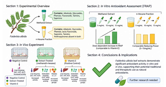
Background and Objective: Oxidative stress, resulting from the accumulation of free radicals, can cause cellular damage and contribute to the development of chronic diseases. Certain medicinal plants exhibit potent antioxidant activity, potentially shielding cells from oxidative harm. This study investigated the phytochemical composition and in vitro and in vivo antioxidant properties of Faidherbia albida (F. albida) leaf extracts, including methanol extract and n-hexane fraction, in albino rats. Materials and Methods: Phytochemical screening was conducted to determine the bioactive constituents of the methanol extract and n-hexane fraction. The in vitro antioxidant activity was assessed using ferric reducing antioxidant power (FRAP), while in vivo antioxidant effects were evaluated in albino rats by measuring ALT, AST, ALP, Catalase (CAT), Superoxide dismutase (SOD), and vitamins A, C, and E. Results: Phytochemical screening detected alkaloids, glycosides, phenols, flavonoids, and tannins in the extracts, while anthraquinones were absent. Flavonoids, saponins, and tannins were present in the methanolic extract but absent in the n-hexane fraction. In vitro analysis showed a dose-dependent increase in FRAP for vitamin E, whereas the n-hexane fraction exhibited comparable reducing power across concentrations of 5-15 mg/mL. In vivo assessments showed significant reductions in ALT, AST, and ALP activities in extract-treated and positive control groups compared with the negative control. Conversely, CAT and SOD activities increased significantly in treated groups. Vitamins A, C, and E levels also increased notably in the treated groups. Conclusion: The findings support the antioxidant potential of F. albida leaf extracts and provide evidence for its traditional use in folklore medicine. The extracts may represent a potential source of natural antioxidants and warrant further investigation for pharmaceutical and therapeutic applications.
| Copyright © 2026 Abubakar et al. This is an open-access article distributed under the Creative Commons Attribution License, which permits unrestricted use, distribution, and reproduction in any medium, provided the original work is properly cited. |
Visual Abstract
 |
INTRODUCTION
The excessive accumulation of free radicals in body tissues can cause oxidative damage to biomolecules and cellular membranes, contributing to chronic diseases such as inflammation, cancer, diabetes, cardiovascular issues, neurodegenerative disorders, and aging1,2. Antioxidants counteract this process by donating or accepting electrons to neutralise free radicals, thus preventing the persistence of unpaired radical states. In this way, antioxidant molecules not only directly interact with reactive radicals but may also undergo structural changes that generate new, less harmful radical intermediates2.
Oxidative stress arises from an imbalance between the excessive production of reactive oxygen species (ROS) and reactive nitrogen species (RNS) and the limited capacity of cellular defence systems to eliminate these reactive intermediates3. Persistent oxidative stress has been implicated in multiple pathological states, including cardiovascular diseases4, chronic inflammatory disorders5, pulmonary diseases6, depression7, and neurodegenerative diseases such as Alzheimer’s and Parkinson’s8. The brain is particularly susceptible to oxidative damage due to its high oxygen consumption, lower antioxidant enzyme activity, abundance of polyunsaturated fatty acids, and the limited regenerative capacity of neurons3. Reactive oxygen species such as superoxide and hydroxyl radicals are potent initiators of lipid peroxidation, a process central to the pathogenesis of many of these diseases9. Given the central role of oxidative stress in disease progression, antioxidants have become increasingly significant as preventive and therapeutic agents.
Medicinal plants are a rich source of bioactive phenolic compounds with therapeutic potential against diabetes, cardiovascular diseases, cancer, and inflammation1,9. Their antioxidant and oxidative damage-protective effects have sparked significant interest in recent years1. One such plant, F. albida (syn. Acacia albida Del.), a member of the Mimosoideae family, is gaining attention for its ethnomedicinal value. Indigenous to Southwest, West, and North Africa, it is locally known as "Gawo" (Hausa) and "Chayski" (Fulfulde). Studies show F. albida has anti-pyretic, anti-inflammatory, anti-diarrheal, and anti-trypanosomal activities in rats1,10. However, limited information exists on its antioxidant properties, especially in both in vitro and in vivo systems. This study investigates phytochemicals and evaluates the antioxidant potential of F. albida leaf methanolic extract and n-hexane fraction in albino rats, providing novel insights into its therapeutic relevance and safety as a natural antioxidant source.
MATERIALS AND METHODS
Study area: The research was conducted at the Biochemistry Research Laboratory, Department of Biochemistry, Abdullahi Fodio University of Science and Technology, Aliero, Nigeria, from March to September 2024 (6 months).
Experimental animals: Albino Wistar rats, sourced from Usmanu Danfodiyo University, Sokoto (July 2024), were acclimatised for two weeks at Kebbi State University’s Animal House before the commencement of the experiment. They were fed standard rodent pellets and water ad libitum.
Collection and Identification of plant sample: Faidherbia albida leaves were collected in April 2024 in Aleiro Town, Aleiro Local Government Area of Kebbi State. The plant was authenticated by a Taxonomist from the Department of Plant Science and Biotechnology, Kebbi State University of Science and Technology, Aleiro. And a Voucher specimen Number (ksusta/psb/h/319) was assigned, and the plant was deposited in the herbarium of the same Department.
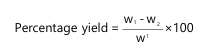
Plant preparation and extraction of F. albida Leaves: Faidherbia albida leaves were washed with clean water and allowed to dry in the shade for two weeks. They were then ground to coarse powder using a grinding machine. 1.5 kg of the powdered leaves were dissolved in 2.5 L of methanol for 72 hrs11. The mixture was filtered using muslin cloth and Whatman No. 1 filter paper, and the filtrate was evaporated using an oven at 45°C. The dried extract was stored in an airtight container and kept in a refrigerator at 4°C. Extraction yield was calculated as follows:
 |
Where:
| w1 | = | Sample weight initially placed in the thimble | |
| w2 | = | Sample weight after dried in the oven |
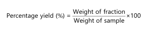
Hexane fractionation of F. albida leaves methanol extract: A 50 g of dried F. albida leaf methanolic extract was redissolved in water and partitioned with n-hexane (50:50 v/v) in a separating funnel12. The mixture was shaken, allowed to separate, and the hexane layer was collected and evaporated to dryness. The remaining water layer was dried to yield an aqueous extract.
 |
Phytochemical screening of F. albida Leaves’ methanolic and hexane fractions
Detection of alkaloids: A 0.2 g extract was boiled with 5 mL of 2% HCl for 20 min, filtered, and 1 mL filtrate was treated with Wagner’s reagent (2 drops). A reddish-brown precipitate formed, indicating alkaloids13.
Detection of flavonoids: A 0.2 g extract was dissolved in 1 mL of 10% NaOH; yellow colour indicates flavonoids13.
Detection of tannins: A 2 mL extract treated with 5% FeCl3 gave a dark green colour, indicating tannins13.
Detection of steroids: A 0.5 g extract was dissolved in 2 mL chloroform. Concentrated H2SO4 layered underneath formed a reddish-brown interface, indicating steroids12.
Detection of saponins: A 0.5 g extract was shaken vigorously with 2.5 mL water. Persistent froth lasting several minutes indicated saponins12.
Detection of dlycosides: A 0.5 g extract was heated with 2.5 mL 50% H2SO4 for 15 min, neutralised with 10% NaOH, then boiled with Fehling's solution. Brick-red precipitate indicated glycosides12.
Detection of phenols: A 1 mL extract treated with 10% FeCl3 gave a blue-green colour, indicating phenolic compounds12.
In-vitro antioxidant activity
Determination of antioxidant potential by FRAP assay: The FRAP of the extracts was determined using the potassium ferricyanide-ferric chloride method described by Oyaizu14. Standard vitamin E was used as a positive reference control. About 2 mL of extract and standard drug vitamin C at various concentrations (250, 500 and 750) was added to 2.5 mL of potassium ferricyanide in separate test tubes, and the mixture was incubated at 50°C for 20 min. Then 2.5 mL of trichloroacetic acid was added to the mixture, and then centrifuged at 650×g for 10 min. To 2.5 mL of the supernatant, 2.5 mL of distilled water and 0.5 mL ferric chloride were added. The absorbance was read at 700 nm using a spectrophotometer. Higher absorbance indicates greater reducing capacity, which was calculated as follows.
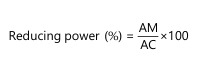 |
Where
| AM | = | Absorbance of reaction mixture | |
| AC | = | Absorbance of control mixture (distilled water) |
In vivo antioxidant assay
Induction of oxidative stress: Oxidative stress was induced in rats via intraperitoneal (i.p.) injection of CCl4 diluted in liquid paraffin 8% v/v at a single dose of 1.25 mL/kg body weight (equivalent to 0.25 mL CCl4 kg)15. Following induction, animals were randomly divided into 5 experimental groups n = 4 and administered treatments once daily via oral gavage p.m. for 7 consecutive days at a dosage volume of 10 mL/kg body weight, as outlined in Table 1.
Determination of lipid peroxidation (Malondialdehyde) level: Lipid peroxidation was determined by measuring the level of malondialdehyde (MDA) spectrophotometrically, as described by Wallin et al.16. Briefly, 0.1 mL of serum was mixed with 0.9 mL of distilled water, followed by the addition of 0.5 mL of 25% trichloroacetic acid and 0.5 mL of 1% thiobarbituric acid in 0.3% NaOH. The mixture was boiled for 40 min in a water bath and then cooled in cold water. Subsequently, 0.1 mL of 20% sodium dodecyl sulfate was added to the cooled solution and mixed properly. The absorbance was then measured at wavelengths 532 nm and 600 nm against a blank.
| Table 1: | Gross composition of solvent extracted processed soyabean based diets | |||
| Group | Treatment |
| Group 1 | Neither induced nor treated, served as the normal control |
| Group 2 | CCl4-induced rats but untreated, served as negative control |
| Group 3 | CCl4-induced rats and treated with vitamin C (5 mg/kg), served as standard control |
| Group 4 | CCl4-induced rats and treated with methanol extracts (400 mg/kg), served as treatment group |
| Group 5 | CCl4-induced rats and treated with N-hexane fraction (400 mg/kg), served as treatment group |
| The extract was administered to the animals orally. The rats were sacrificed on the seventh day of the experiment. Blood samples were collected in heparinised bottles for antioxidant analysis | |
Assay for superoxide dismutase (SOD) activity: SOD activity was determined using the adrenaline auto-oxidation inhibition method of Xin et al.17. A 0.01 g sample of adrenaline was dissolved in 17 mL of distilled water. Then, 0.1 mL of serum and 0.9 mL of phosphate buffer (pH 7.8) were taken in triplicate in 2.5 mL buffer. Next, 0.3 mL of adrenaline solution was added and mixed inside the cuvette. Absorbance was measured at 480 nm at 30 sec intervals, five times. The rate of change in absorbance was used to determine superoxide dismutase activity using this equation:
 |
Where:
ABS = Absorbance
Assay for catalase activity: The activity of catalase was assayed according to the method of Aebi18. To 2 mL of hydrogen peroxide, 2.5 mL of phosphate buffer was added to a beaker, followed by 0.5 mL of the sample. Then, a 1 mL portion of the reaction mixture was added to 2 mL of dichromate-acetic acid reagent. The absorbance was read at 570 nm at one-minute intervals, four times. Catalase activity was calculated using the following equation:
 |
Where:
ABS = Absorbance
Determination of reduced glutathione (GSH): GSH was determined based on the method of Jollow et al.19. A 0.1 mL sample was mixed with 0.9 mL of distilled water, followed by the addition of 1 mL of 2% sodium sulphate. Then, 0.02 mL of 20% lithium sulphate and 0.2 mL of 20% Na2CO3 were added, and the mixture was shaken vigorously. Next, 0.2 mL of phospho-18-tungstic acid was added. The absorbance was measured at 650 nm:
 |
Determination of alkaline phosphatase (ALP): Alkaline phosphatase activity was estimated using the method of Ramnik20. Serum (0.02 mL) was placed into a labelled test tube, and 1 mL of the alkaline phosphatase working reagent was added. The mixture was mixed, and the initial absorbance was read at 405 nm at zero min. A timer was started, and absorbance readings were taken at 1, 2, and 3 min at 405 nm.
Determination of aspartate aminotransferase (AST): The AST catalytic activity was determined by the method of Reitman and Frankel21. Appropriate volumes of the required solutions were dispensed into well-labelled test tubes. Distilled water (0.1 mL) and sample (0.1 mL) were pipetted into blank and sample test tubes, respectively. Reagent 1 (0.5 mL) was added to both test tubes, mixed, and incubated for 30 min at 37°C. Then, 0.5 mL of reagent 2 was added to both test tubes, mixed, and allowed to stand for 20 min at 25°C. Next, 0.4 M NaOH was added to both test tubes. The contents were mixed, and absorbance was read against the blank after 5 min at 546 nm. AST activity in the serum was obtained from a standard curve.
Determination of alanine aminotransferase (ALT): The ALT activity was determined by the method of Reitman and Frankel21. Appropriate volumes of the required solution were dispensed into well-labelled test tubes. Distilled water (0.1 mL) and sample (0.1 mL) were pipetted into blank and sample test tubes, respectively. Reagent 1 (0.5 mL) was added to both test tubes, mixed, and incubated for 30 min at 37°C. Then, 0.5 mL of reagent 2 was added to both test tubes, mixed, and allowed to stand for 20 min at 25°C. Next, 0.4 M NaOH was added. The contents were mixed, and absorbance was read against the blank after 5 min at 546 nm. ALT activity in the serum was obtained from a standard curve.
Determination of vitamin A: Vitamin A concentration was determined using the method of Laurence and Sobel22. One mL of serum was pipetted into a test tube, and 1 mL of 95% ethanol was added. The contents were mixed by tapping. For saponification, 1 mL of 1 N KOH in 90% ethanol was added, mixed, and incubated at 60°C for 20 min. Two mL of petroleum ether (analytical reagent) was added, and the tube was shaken for 10 min, then centrifuged for 30 sec. The supernatant petroleum ether was aspirated using a fine-tipped dropper and transferred to a test tube. The extraction was repeated with another 2 mL of petroleum ether, shaking for 5 min. Carotene was determined at 440 nm; the petroleum ether extract was transferred to a 13 mm cuvette, and the volume was adjusted to 4.0 mL. Absorbance was read at 440 nm using Filter PC-4:
 |
Determination of vitamin C: Vitamin C concentration was determined using the method of Laurence and Sobel22. One mL of serum was pipetted into a test tube, and 1 mL of 95% ethanol was added. The contents were mixed by tapping. For saponification, 1 mL of 1 N KOH in 90% ethanol was added, mixed, and incubated at 60°C for 20 min. Two mL of petroleum ether (analytical reagent) was added, and the tube was shaken for 10 min, then centrifuged for 30 sec. The supernatant petroleum ether was aspirated using a fine-tipped dropper and transferred to a test tube. The extraction was repeated with another 2 mL of petroleum ether, shaking for 5 min. The petroleum ether extract was transferred to a 13 mm cuvette, and the volume was adjusted to 4.0 mL. Absorbance was read at 440 nm using Filter PC-4:
 |
Determination of vitamin E: This method is based on the reduction of ferric ions by serum vitamin E to ferrous ions, which form a red colour with dipyridyl, measured spectrophotometrically at 520 nm23. Three test tubes were labelled sample, standard, and blank. To the sample test tube, 1.5 mL of serum was added. A 1.5 mL standard solution was added to the standard test tube. Ethanol (1.5 mL) was added to the sample and blank test tubes. Then, 1.5 mL of distilled water was added to the standard and blank test tubes. Xylene (1.5 mL) was added to all three test tubes. The contents were mixed and centrifuged for 10 min. One mL of the xylene layer was carefully pipetted into another set of test tubes, and 1 mL of α-dipyridyl reagent was added. The contents were mixed, and the absorbance of the sample and standard was read against the blank at 460 nm. After 5 min, 0.33 mL of ferric chloride solution was added. The contents were mixed, and the absorbance was read at 520 nm.
Standard composition: D-α-tocopherol, 10 mg/L in ethanol:
 |
Data analysis: The data generated from the study are presented as Mean±Standard Error of Mean (SEM). One-way Analysis of Variance (ANOVA) was performed, and statistical differences between means were determined using Duncan’s Multiple Comparison Test, via Statistical Package for Social Sciences (SPSS)24 version 20. Values with p<0.05 were considered statistically significant.
Ethical statement: All experimental procedures involving animals were reviewed and approved by the University Research Ethics Committee (UREC), Abdullahi Fodio University of Science and Technology, Aliero (approval number: KSUSTA/DVC R&I/RECC/007), and were conducted in accordance with institutional guidelines for the care and use of laboratory animals.
RESULTS
Percentage yield: The extraction of F. albida leaves with methanol yielded 5.8%, while the hexane fraction yielded 1.4%. The extract is light green in colour, sour in taste, has a nice smell, and a gummy texture, and is water-soluble.
Qualitative phytochemical analysis of methanol and hexane fractions from F. albida leaves: The qualitative phytochemical screening of F. albida leaf methanolic and hexane fractions is presented in Table 2. The methanolic extract revealed the presence of alkaloids, steroids, saponins, glycosides, flavonoids, tannins, and phenols. The hexane fraction revealed the presence of alkaloids, steroids, saponins, glycosides, flavonoids, and tannins, while anthraquinones and phenols were not detected.
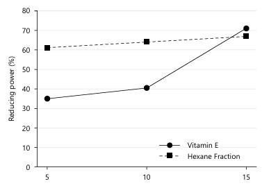
FRAP of F. albida methanol extract and hexane fraction: The FRAP of F. albida methanol extract is presented in Fig. 1. The result demonstrates a dose-dependent increase in FRAP for vitamin E, while F. albida leaf hexane fraction showed similar reducing power at all concentrations (5, 10, and 15 mg/mL).
In vivo antioxidant potentials of F. albida methanol extract and hexane fraction: The antioxidant potential of F. albida leaf hexane fraction is presented in Table 3. The MDA concentration of the negative control was significantly (p<0.05) higher than that of the normal control, positive control, methanol extract, and hexane fraction. However, the positive control and methanol extract showed significant (p<0.05) increases compared to the normal control, whereas there was no significant (p>0.05) difference in the concentration of the hexane fraction compared to the normal control.
The GSH and catalase (CAT) concentrations of the negative control were significantly (p<0.05) lower than those of the normal control, positive control, and all extract-treated groups. There was no significant (p>0.05) difference in GSH concentration between the positive control, methanol extract, and hexane fraction compared to the normal control. The superoxide dismutase (SOD) concentration of the negative control was significantly (p<0.05) lower than that of the normal control, positive control, and all extract-treated groups. There was no significant (p>0.05) difference in SOD concentration between the positive control and hexane fraction compared to the normal control, while there was a significant (p<0.05) decrease in the concentration of the methanol extract compared to the normal control.
The vitamin A concentration of the negative control was significantly (p<0.05) higher than that of the normal control, positive control, and all extract-treated groups. However, there was a significant (p<0.05) decrease in vitamin A concentration of the positive control and hexane fraction compared to the normal control, while there was a significant (p<0.05) increase in the concentration of the methanol extract compared to the normal control. The vitamin C concentration of the negative control was significantly (p<0.05) lower than that of the normal control, positive control, and all extract-treated groups. However, there was a significant (p<0.05) increase in vitamin C concentration of the positive control and methanol extract compared to the normal control, while the hexane fraction was significantly (p<0.05) lower than the normal control.
|
| Table 2: | Qualitative phytochemical profile of methanol extract and hexane fractions from F. albida leaves | |||
| Phytochemicals | Methanol extract | Hexane fraction |
| Flavonoids | + | - |
| Tannins | + | - |
| Steroids | + | + |
| Saponins | + | - |
| Glycosides | + | + |
| Alkaloids | + | + |
| Anthraquinone | - | - |
| Phenols | + | + |
| +: Detected and =: Not detected | ||
The vitamin E concentration of the negative control was significantly (p<0.05) higher than that of the normal control, positive control, and all extract-treated groups. However, there was a significant (p<0.05) decrease in vitamin E concentration of the positive control and methanol extract compared to the normal control, and a significant (p<0.05) decrease in the hexane fraction compared to the normal control.
The ALT concentration of the negative control was significantly (p<0.05) higher than that of the normal control, positive control, and methanol extract, but significantly (p<0.05) lower than that of the hexane fraction. The ALT concentration of the positive control, methanol extract, and hexane fraction was significantly (p<0.05) higher than that of the normal control. The AST concentration of the negative control was significantly (p<0.05) higher than that of the normal control, positive control, and all extract-treated groups. However, there was a significant (p<0.05) increase in AST concentration of the positive control, methanol extract, and hexane fraction compared to the normal control. The ALP concentration of the negative control was significantly (p<0.05) higher than that of the normal control and hexane fraction. There was no significant (p>0.05) difference in the concentration of the negative control compared to the positive control and the methanol extract. The positive control, methanol extract, and hexane fraction were significantly (p<0.05) higher than the normal control.
DISCUSSION
Phenolic compounds and their derivatives are known for their antioxidative and free radical-scavenging properties. The antioxidant activity of medicinal plants has been linked to their phytochemical compositions25. The presence of phytoconstituents such as phenols, flavonoids, tannins, and saponins in the methanolic extract of F. albida leaves may strongly contribute to its traditional use in managing diseases. Total phenols and flavonoids have demonstrated modulatory effects against lipid peroxidation involved in atherogenesis and carcinogenesis25. Tannins are polyphenolic compounds with antidiuretic and antidiarrheal properties25. Saponins have shown potential therapeutic activities like antifungal, antibacterial, and antioxidant agents25. The polyphenols present in F. albida leaf extract may synergistically enhance its overall antioxidant potential. Synergy among phenolic compounds (such as flavonoids, phenolic acids, and tannins) occurs when their combined free radical scavenging activity exceeds the sum of their individual effects. Recent studies show that polyphenolic mixtures can interact through complementary mechanisms, including hydrogen donation, metal chelation and regeneration of one another after oxidation, resulting in stronger antioxidant responses25,26. Evidence from similar medicinal plants demonstrates that synergistic interactions between polyphenols significantly boost DPPH, FRAP and ABTS activities, supporting the likelihood that such cooperative effects contribute to the strong antioxidant properties observed in F. albida leaf extract27,28. Thus, the combined action of multiple polyphenols is likely a key factor underpinning the potent antioxidant capacity of F. albida.
| Table 3: | Antioxidant potentials of F. albida methanol extract and hexane fraction | |||
| Treatment | Normal control | Negative control (CCL4) |
Positive Control Vit C (5 mg/kg) |
Methanol extract (400 mg/kg) |
Hexane fraction (400 mg/kg) |
| MDA (μmole/L) | 104.48±1.34a | 245.29±1.54c | 119.23±1.11b | 116.02±0.98b | 106.62±0.57a |
| GSH (mg/dL) | 123.34±0.25b | 40.23±0.39a | 120.81±0.36b | 123.78±3.80b | 120.70±9.47b |
| CAT (unit/mL) | 1.93±0.12b | 0.83±0.04a | 1.62±0.09b | 1.86±0.11b | 1.95±0.20b |
| SOD (unit/mL) | 3.76±0.23c | 1.18±0.10a | 3.37±0.25c | 2.20±0.11b | 3.61±0.28c |
| VIT A (mg/dL) | 69.60±0.70b | 102.67±0.21d | 59.76±0.41a | 76.93±0.29c | 60.94±0.36a |
| VIT C (μmol/L) | 797.51±1.22c | 41.56±0.80a | 922.18±1.22d | 1136.92±1.66e | 549.06±3.61b |
| VIT E (mg/dL) | 77.39±0.19c | 124.89±0.25e | 53.86±0.37a | 106.65±0.37d | 71.03±0.25b |
| ALT (U/L) | 12.22±0.22a | 19.50±0.73c | 17.17±0.27b | 18.24±0.29b | 28.14±0.23d |
| AST (U/L) | 79.40±0.63a | 147.46±0.63e | 115.52±0.63c | 128.08±0.46d | 102.96±0.92b |
| ALP (U/L) | 31.93±3.35a | 83.75±2.73c | 71.90±0.14bc | 88.32±5.75c | 56.16±14.61b |
| Values are presented as Mean±SEM (n = 3). Values with similar superscripts in rows are not significantly different (p>0.05), as analysed using One-Way ANOVA followed by Duncan’s multiple comparison test with SPSS version 20.0. Abbreviations: SOD: Superoxide dismutase, CAT: Catalase, GSH: Reduced glutathione, MDA: Malondialdehyde, VIT A: Vitamin A, VIT C: Vitamin C, VIT E: Vitamin E, ALT: Alanine transaminase, AST: Aspartate transaminase and ALP: Alkaline phosphatase | |||||
In this study, the iron-reducing capacity of vitamin E and the hexane fraction was estimated by their ability to reduce the Fe3+ ferricyanide complex to the Fe2+ form by donating an electron. The reducing ability of vitamin E was in the range of 35-70% with increased doses of 5, 10, and 15 mg/mL (Fig. 1), while the F. albida leaf hexane fraction showed similar reducing powers at all concentrations (5, 10, and 15 mg/mL). All the compounds showed good reducing power capacity. The results are consistent with earlier findings29 and align with more recent studies demonstrating strong antioxidant activity in F. albida attributable to its rich polyphenolic content. Recent research confirms that F. albida extracts exhibit high DPPH and FRAP activities, strongly correlated with phenolic and flavonoid levels27,30. Polyphenols act as potent antioxidants by donating electrons or hydrogen atoms to neutralize free radicals, a mechanism widely reported in phytochemical studies25,26,28. These observations strengthen our conclusion that the antioxidant and reducing power capacities of F. albida are largely mediated by its polyphenolic constituents.
The results of lipid peroxidation (Table 3) revealed a significant (p<0.05) decrease in tissues for the groups treated with methanol extract (400 mg/kg) and hexane fraction (400 mg/kg) compared to the negative control, while the positive control (vitamin C, 5 mg/kg) showed a significant (p<0.05) decrease compared to the normal control group. Lipid peroxidation is an auto-catalytic, free radical-mediated, destructive process whereby polyunsaturated fatty acids in cell membranes undergo degradation to form lipid hydroperoxides31. These antioxidants may act in a complementary and synergistic manner to provide better protection against ROS25,26. In the present study, the reversal of lipid peroxidation activity by the plant extract may be attributed to carbon tetrachloride (CCl4), a liver toxicant, whose metabolites, such as trichloromethyl radical (CCl3•) and trichloromethyl peroxyl radical (CCl3O2•) are involved in the pathogenesis of liver and kidney damage15. Superoxide dismutase (SOD), an important intracellular antioxidant enzyme present in all aerobic cells, has an antitoxic effect against superoxide anion17.
The non-enzymatic antioxidant glutathione is one of the most abundant tripeptides present in the liver. Its functions are mainly concerned with removing free radical species such as hydrogen peroxide, superoxide radicals, and alkoxy radicals, maintaining membrane protein thiols, and serving as a substrate for glutathione peroxidase and GST19. The body has evolved a complex defence strategy to minimise the damaging effects of various oxidants. CAT is an enzymatic antioxidant widely distributed in all animal tissues, decomposing hydrogen peroxide and protecting tissues from highly reactive hydroxyl radicals18. Similar research indicates that in an in vivo model, Ruta graveolens L. reduces oxidative stress by decreasing reactive hydroxyl radical levels and increasing antioxidant enzyme activities such as SOD, catalase, and GPX in the liver and heart of hypercholesteromic rats29. Beslo et al.31 also indicated in vitro that Ruta graveolens L. extract scavenges hydroxyl radicals and inhibits lipid peroxidation.
Ascorbic acid exerts its protective effects primarily through its well-established free radical scavenging mechanism32. Recent studies further support that ascorbic acid donates electrons to neutralize reactive oxygen species, thereby preventing lipid peroxidation and oxidative damage in biological systems32,33. Its role as a reducing agent also enables the regeneration of other antioxidants, such as vitamin E, enhancing overall antioxidant defense32,33. These findings reinforce the view that the cytoprotective properties of ascorbic acid are closely linked to its ability to quench free radicals and maintain redox homeostasis.
The groups treated with methanol extract (400 mg/kg) and hexane fraction (400 mg/kg) showed a significant (p<0.05) decrease in ALT, AST, and ALP activities compared to the control group and positive control group (Table 3). We suggest that this could be due to natural healing that possibly occurred hours after extract treatment, confirming that liver cells are capable of regenerating naturally20,21, especially when the toxic agent is eliminated. This also indicates that using a high dosage of CCl creates an ideal hepatotoxicity model in rats, which may persist for a longer time.
CONCLUSION
The present study set out to characterise the phytochemical composition and antioxidant potential of F. albida leaf methanol extract and its hexane fraction, in both in vitro and in vivo systems. Phytochemical screening confirmed the presence of alkaloids, glycosides, phenols, flavonoids, saponins, and tannins, and both extracts exhibited dose-dependent in vitro reducing power and significantly restored in vivo antioxidant enzyme activities (CAT, SOD), vitamin status, and liver biomarkers (ALT, AST, ALP) following CCl4-induced oxidative stress. These findings address the study’s objective by providing direct evidence, in both in vitro and in vivo systems that supports the traditional folkloric use of F. albida and identifies it as a promising source of natural antioxidant compounds warranting further pharmaceutical investigation.
SIGNIFICANCE STATEMENT
This study highlights the antioxidant potential of Faidherbia albida leaf extracts through in vitro and in vivo assessments. The extracts improved antioxidant enzyme activities, vitamin levels, and liver function markers in albino rats, supporting their potential as a source of natural antioxidants. These findings provide a scientific basis for the traditional medicinal use of F. albida and encourage further research into its therapeutic applications.
- Muscolo, A., O. Mariateresa, T. Giulio and R. Mariateresa, 2024. Oxidative stress: The role of antioxidant phytochemicals in the prevention and treatment of diseases. Int. J. Mol. Sci., 25.
- Ofoedu, C.E., L. You, C.M. Osuji, J.O. Iwouno and N.O. Kabuo et al., 2021. Hydrogen peroxide effects on natural-sourced polysacchrides: Free radical formation/production, degradation process, and reaction mechanism-A critical synopsis. Foods, 10.
- Olufunmilayo, E.O., M.B. Gerke-Duncan and R.M.D. Holsinger, 2023. Oxidative stress and antioxidants in neurodegenerative disorders. Antioxidants, 12.
- Dubois-Deruy, E., V. Peugnet, A. Turkieh and F. Pinet, 2020. Oxidative stress in cardiovascular diseases. Antioxidants, 9.
- Leyane, T.S., S.W. Jere and N.N. Houreld, 2022. Oxidative stress in ageing and chronic degenerative pathologies: Molecular mechanisms involved in counteracting oxidative stress and chronic inflammation. Int. J. Mol. Sci., 23.
- Bezerra, F.S., M. Lanzetti, R.T. Nesi, A.C. Nagato and C. Pecli e Silva et al., 2023. Oxidative stress and inflammation in acute and chronic lung injuries. Antioxidants, 12.
- Tayeb, A.E.K.A., V. Poinsignon, K. Chappell, J. Bouligand, L. Becquemont and C. Verstuyft, 2023. Major depressive disorder and oxidative stress: A review of peripheral and genetic biomarkers according to clinical characteristics and disease stages. Antioxidants, 12.
- Ionescu-Tucker, A. and C.W. Cotman, 2021. Emerging roles of oxidative stress in brain aging and Alzheimer's disease. Neurobiol. Aging, 107: 86-95.
- Martemucci, G., C. Costagliola, M. Mariano, L. D’andrea, P. Napolitano and A.G. D’Alessandro, 2022. Free radical properties, source and targets, antioxidant consumption and health. Oxygen, 2: 48-78.
- Tijani, A.Y., M.O. Uguru, O.A. Salawu, A. Abubakar, N.O. Onyekwelu and J.A. Akingbasote, 2009. Effect of Faidherbia albida on some biochemical parameters of rats infected with Trypanosoma brucei brucei. Afr. J. Pharm. Pharmacol., 3: 26-30.
- Dupont, É., P. Falardeau, S.A. Mousa, V. Dimitriadou, M.C. Pepin, T. Wang and M.A. Alaoui-Jamali, 2002. Antiangiogenic and antimetastatic properties of Neovastat (Æ-941), an orally active extract derived from cartilage tissue. Clin. Exp. Metastasis, 19: 145-153.
- Hammadi, R., N. Kúsz, C.Z. Dávid, Z. Behány and L. Papp et al., 2021. Ingol and ingenol-type diterpenes from Euphorbia trigona Miller with keratinocyte inhibitory activity. Plants, 10.
- Harborne, J.B., 1973. Phytochemical Methods: A Guide to Modern Techniques of Plant Analysis. 1st Edn., Springer, Dordrecht, Netherlands, ISBN: 978-0-412-23050-9, Pages: 278.
- Oyaizu, M., 1986. Studies on products of browning reaction: Antioxidative activities of products of browning reaction prepared from glucosamine. Jpn. J. Nutr. Diet., 44: 307-315.
- Chougale, A.D., S.N. Panaskar, P.M. Gurao and A.U. Arvindekar, 2007. Optimization of alloxan dose is essential to induce stable diabetes for prolonged period. Asian J. Biochem., 2: 402-408.
- Wallin, B., B. Rosengren, H.G. Shetzer and G. Camejo, 1993. Lipoprotein oxidation and measurement of thiobarbituric acid reacting substances formation in a single microtiter plate: Its use for evaluation of antioxidants. Anal. Biochem., 208: 10-15.
- Xin, Z., D.F. Waterman, R.W. Hemken and R.J. Harmon, 1991. Effects of copper status on neutrophil function, superoxide dismutase, and copper distribution in steers. J. Dairy Sci., 74: 3078-3085.
- Aebi, H., 1974. Catalase. In: Methods of Enzymatic Analysis, Bergneyer, H. (Ed.), Academic Press, Weinham, New York, pp: 673-684.
- Jollow, D.J., J.R. Mitchell, N. Zampaglione and J.R. Gillette, 1974. Bromobenzene-induced liver necrosis. Protective role of glutathione and evidence for 3,4-bromobenzene oxide as the hepatotoxic metabolite. Pharmacology, 11: 151-169.
- Ramnik, S., 2009. Medical Laboratory Technology: Methods and Interpretations. Jaypee Brothers Medical Publishers Pvt. Limited, New Delhi, India, pp: 1694.
- Reitman, S. and S. Frankel, 1957. A colorimetric method for the determination of serum glutamic oxalacetic and glutamic pyruvic transaminases. Am. J. Clin. Pathol., 28: 56-63.
- Laurence, P.A. and A.E. Sobel, 1953. Changes in serum vitamin A level during the human menstrual cycle. J. Clin. Endocrinol. Metab., 13: 1192-1200.
- Jargar, J.G., S.H. Hattiwale, S. Das, S.A. Dhundasi and K.K. Das, 2012. A modified simple method for determination of serum α-tocopherol (vitamin E). J. Basic Clin. Physiol. Pharmacol., 23: 45-48.
- Field, A., 2018. Discovering Statistics Using IBM SPSS Statistics. SAGE Publications, Thousand Oaks, California, USA, ISBN: 9781526419514, Pages: 1104.
- Feng, Y., C. Jin, S. Lv, H. Zhang, F. Ren and J. Wang, 2023. Molecular mechanisms and applications of polyphenol-protein complexes with antioxidant properties: A review. Antioxidants, 12.
- Swallah, M.S., H. Yu, C. Piao, H. Fu, Z. Yakubu and F.L. Sossah, 2021. Synergistic two-way interactions of dietary polyphenols and dietary components on the gut microbial composition: Is there a positive, negative, or neutralizing effect in the prevention and management of metabolic diseases? Curr. Protein Pept. Sci., 22: 313-327.
- Sarr, A., S.I.M. Dieng, A.I. Mbaye, M.L. Dieng and K. Diatta et al., 2024. Phytochemical screening and nutritional assessment of bioactive compounds in two Senegalese medicinal plants: Faidherbia albida Del. and Ziziphus mauritiana Lam. Asian J. Res. Med. Pharm. Sci., 13: 202-209.
- Lv, Q.Z., J.T. Long, Z.F. Gong, K.Y. Nong and X.M. Liang et al., 2021. Current state of knowledge on the antioxidant effects and mechanisms of action of polyphenolic compounds. Nat. Prod. Commun., 16.
- Huang, D., B. Ou and R.L. Prior, 2005. The chemistry behind antioxidant capacity assays. J. Agric. Food Chem., 53: 1841-1856.
- Abdelwahab, S.I., M.M.E. Taha, A.S. Al-Zubairi, A. Syahida and L. KaHeng et al., 2024. Anti-inflammatory and antioxidant properties of bark and fruit extracts of Faidherbia albida (Delile) A. Chev: A perspective from bio-prospecting assays to scientometric approach. AIMS Mol. Sci., 11: 262-276.
- Bešlo, D., N. Golubić, V. Rastija, D. Agić, M. Karnaš, D. Šubarić and B. Lučić, 2023. Antioxidant activity, metabolism, and bioavailability of polyphenols in the diet of animals. Antioxidants, 12.
- Popovic, L.M., N.R. Mitic, D. Miric, B. Bisevac, M. Miric and B. Popovic, 2015. Influence of vitamin C supplementation on oxidative stress and neutrophil inflammatory response in acute and regular exercise. Oxid. Med. Cell. Longevity, 2015.
- Mandl, J., A. Szarka and G. Bánhegyi, 2009. Vitamin C: Update on physiology and pharmacology. Br. J. Pharmacol., 157: 1097-1110.
How to Cite this paper?
APA-7 Style
Abubakar,
S.J., Sani,
I., Hussain,
N., Nasir,
A., Usman,
S. (2026). Antioxidant Potential of Faidherbia albida Leaf Extracts: In vitro and in vivo Studies in Albino Rats. Trends in Biological Sciences, 2(4), 451-462. https://doi.org/10.21124/tbs.2026.451.462
ACS Style
Abubakar,
S.J.; Sani,
I.; Hussain,
N.; Nasir,
A.; Usman,
S. Antioxidant Potential of Faidherbia albida Leaf Extracts: In vitro and in vivo Studies in Albino Rats. Trends Biol. Sci 2026, 2, 451-462. https://doi.org/10.21124/tbs.2026.451.462
AMA Style
Abubakar
SJ, Sani
I, Hussain
N, Nasir
A, Usman
S. Antioxidant Potential of Faidherbia albida Leaf Extracts: In vitro and in vivo Studies in Albino Rats. Trends in Biological Sciences. 2026; 2(4): 451-462. https://doi.org/10.21124/tbs.2026.451.462
Chicago/Turabian Style
Abubakar, Sufiyanu, Jiga, Ibrahim Sani, Naveed Hussain, Abduljabbar Nasir, and Safiya Usman.
2026. "Antioxidant Potential of Faidherbia albida Leaf Extracts: In vitro and in vivo Studies in Albino Rats" Trends in Biological Sciences 2, no. 4: 451-462. https://doi.org/10.21124/tbs.2026.451.462

This work is licensed under a Creative Commons Attribution 4.0 International License.