
Bioactive Constituents, Hypoglycemic, Antioxidative and Hypolipidemic Potentials of Tridax procumbens Plant Leaf Diets in Experimental Rats
-
Folake Lucy Oyetayo


Department of Biochemistry, Ekiti State University Ado-Ekiti, Nigeria
Elijah Olalekan OdesanmiDepartment of Biochemistry, Ekiti State University Ado-Ekiti, Nigeria
Eniola Elizabeth AkomolafeDepartment of Biochemistry, Ekiti State University Ado-Ekiti, Nigeria
Sheriff Ayodeji AfolabiDepartment of Biochemistry, Ekiti State University Ado-Ekiti, Nigeria
| Received 28 Jan, 2026 |
Accepted 04 Aug, 2026 |
Published 31 Dec, 2026 |
Background and Objective: The Tridax procumbensplant also known as tridax daisy, a herbaceous plant with white and yellow flowers Flowers and leaves with serrated margins found in tropical and sub-tropical areas of the world grows with annual crops, along roadsides, and waste areas. Trado-medically, the leaves of the Tridax procumbensplant are used in the treatment of several ailments: Malaria, high blood pressure and diabetes. However, limited data exist on the scientific basis for these folkloric claims. This study aimed to evaluate the phytochemical bioactives in the leaf of the Tridax procumbens plant, to confirm and document the scientific basis for the use of the leaf as an hypoglycemic agent. Materials and Methods: The Tridax procumbensleaf was screened for the presence of phytochemical bioactives and phenolics were identified and quantified using High-Performance Liquid Chromatography with Diode-Array Detection (HPLC-DAD). Hyperglycemia induced streptozotocin (STZ) which resulted in significant (p<0.05) increase in blood glucose levels in male experimental rats was reversed by the inclusion of Tridax procumbens leaf in their diets with a concomitant significant increase (p<0.05) in antioxidant enzymes activities and inhibition of MDA production. Futhermore, there was a significant decrease (p<0.05) in tissue and serum total cholesterol and the activities of pancreatic amylase and intestinal glucosidase were inhibited compared to those of the diabetic untreated control. Results: Phytochemical analysis confirmed the presence of saponins, flavonoids, tannins, alkaloids, and phenols in the leaf. HPLC-DAD revealed phenolics such as quercetin, kaemferol, rutin, chlorogenic acd acid and coumarin. The inclusion of Tridax procumbens leaf in experimental diets led to a significant decrease (p<0.05) in blood glucose levels of hyperglycemic rats, increase in antioxidant enzymes activities, HDL cholesterol level and inhibition of MDA production. Also, there was a significant decrease (p<0.05) in the activities of pancreatic amylase and intestinal glucosidase compared to the diabetic control which signify delay in carbohydrate digestion and eventual diabetes control. Conclusion: The Tridax procumbens plant leaf holds promise as a rich source of an array of phytochemical compounds with significant medicinal potentials which could serve as alternative phytotherapetic agent for the prevention and management of hyperglycemia in diabetes and other oxidative stress related disorders.
| Copyright © 2026 Oyetayo et al. This is an open-access article distributed under the Creative Commons Attribution License, which permits unrestricted use, distribution, and reproduction in any medium, provided the original work is properly cited. |
INTRODUCTION
Diabetes mellitus resulting from defects in insulin secretion by pancreatic β-cell, insulin inactivity, or both leads to hyperglycemia1. This disorder leads to derangement in carbohydrate, protein, lipid metabolism, free radicals and defence mechanisms due to defect in insulin production or action. There are two main types of diabetes: Type 1 and type 2, with type 2 diabetes accounting for nearly 90% of all diabetes cases, with a worldwide prevalence of 537 million2. Two major types of Diabetes mellitus (DM) exists: DM1 and DM 2 of which DM 2 is more prevalent.
It is a global health concern with increasing prevalence, and it poses significant challenges due to its association with various complications, including retina, cardiovascular diseases, kidney failure, and neuropathy3.Treatment of diabetes with synthetic drugs such as voglibose, which acts as inhibitor of carbohydrate metabolizing enzymes has shown a setback such as its gastrointestinal adverse effects4, and hence may not be recommended for long-term use. Also, the fact that about 80 % of the diabetic people are living in low- and middle-income countries5 where cost of living is high and patients may be unable to afford the cost of their medication. Medicinal plants play important roles in traditional medicine and as precursors of modern pharmaceuticals and lead molecules for the treatment of many diseases from accient times.
Tridax procumbens plant also known as coat button or tridax daisy is found in tropical and sub-tropical areas of the world.It belongs to the family Asteraceae,and grows with annual crops, along roadsides, and waste areas. It is herbaceous with white, tubular, yellow flowers with hairs,leaves with serrated margins. Trado-medically, Tridax procumbens leaf is used in the treatment of malaria, gastrointestinal and respiratory infections, high blood pressure and diabetes6. The presence of Phytochemical compounds have been reported in plants to possess various potential health benefiting properties, including antioxidant activities. The plant is rich in crude proteins, crude fibre and carbohydrates. The search for novel plants with medicinal property which will serve as substitutes for often expensive conventional drugs in the developing world is imperative. The Present study aimed to determine the effects of Tridax procumbens leaf-inclusive diets on the activities of antioxidative stress markers, carbohydrate metabolizing enzymes and lipid profile in hyperglycemia-induced rats. To identify the phenolic constituents of Tridax procumbens plant leaf, we also sought to characterize them with High performance Liquid chromatography coupled diode array detector (HPLC-DAD) and to provide a scientific basis for the folkloric medicinal claims attributed to the plant.
MATERIALS AND METHODS
Study area and duration: The study was carried out in Iworoko of Irepodun/Ifelodun local government Area with coordinates 7°43’ 53” N Latitude 5°15’48” E Longitude between March 2021 and May 2022.
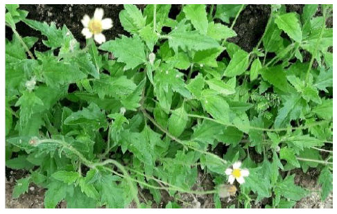
Sample collection and preparation: Tridax procumbens leaves (Fig. 1) were collected from Iworoko Area, Ado-Ekiti, Ekiti State, Nigeria and authenticated by Mr. F.O. Omotayo a plant scientist at Ekiti State University, Herbarium, Ado Ekiti, Ekiti State. Leaves were air-dried for 8 days and then powdered and kept in an air-tight container before analysis.
Phytochemical screening of Tridax procumbens leaf
Test for flavonoid: Five millilitres of dilute ammonia solution was added to 2 mL of the extract. This was followed by the addition of 2 mL concentrated H2SO4. A yellow coloration was observed and the yellow colouration disappeared on standing8.
Test for saponin: The ability of saponins to produce frothing in aqueous solution was used as a screening test for the compound. Exactly 1 mL of the extract was shaken with 2 mL of distilled water in a test tube, and subsequent manifestation of frothing was carefully observed9.
|
Test for tannin: Five milliliters of the extract was stirred with 5 mL distilled water, and the mixture was filtered. The filtrate was treated with ferric chloride, and the presence of a black-green precipitate showed the presence of tannins10.
Test for phlobatannin: Five milliliters of the extract was boiled with 5% aqueous HCl. The absence of a red precipitate indicated the absence of phlobatannins11.
Test for alkaloid: Five grams of the powdered sample were weighed into 250 mL beaker. Then 100 mL of 10% acetic acid in ethanol was added. The mixture was covered and allowed to stand for 4 hrs. This was then filtered and the extract concentrated on a water bath to 1/4 of the original volume. Thereafter, concentrated ammonium hydroxide was added drop wisely until precipitation was completed. The solution was then allowed to settle and the precipitate collected, washed with diluted ammonium hydroxide and filtered. The residue that was dried and weighed was alkaloid11.
Test for steroid: Two milliliters of acetic acid was added to 0.5 mL of the extract. Two milliliters of concentrated H2SO4 was there after added. The presence of a violet to blue colour indicated the prescence of steroids11.
Test for terpenoid: Five milliliters of the extract was mixed with 2 mL chloroform. Three milliliters of concentrated H2SO4 was carefully added to the solution to form a thin layer. The absence of a reddish-brown coloration at the interface gave a negative result for terpenoids11.
Test for cardiac glycoside-salkowski test: About 2 mL of the extract was hydrolysed in 2 mL of HCL solution and neutralized with equal amount of sodium hydroxide solution. Few drops of Fehling’s solution was added. Red precipitates indicated the presence of glycosides12.
Animal management: Thirty-six adult male albino rats of the Wistar strain weighing around 180-200 g were purchased from the animal house of the College of Medicine, Ekiti State University, Ado-Ekiti, Ekiti State, Nigeria. The animals were randomly distributed in six cages of six animals per group under an ambient temperature of 25±2°C, 12±1 hrs light and dark schedule. Rats were acclimatized for 12 days and fed with commercially available rat pellets and water ad libitum. The experimental protocol was approved by the Ekiti State University Office of Research and Development.
| Table 1: | Composition (g/kg) of diets of rats fed control and Tridax procumbens diets | |||
| Groups | 1 | 2 | 3 | 4 | 5 | 6 |
| Skimmed milk | 40 | 40 | 40 | 39.2 | 38.3 | 35.83 |
| Corn Starch | 46 | 46 | 46 | 44.86 | 43.83 | 40.53 |
| Oil | 10 | 10 | 10 | 9.94 | 9.87 | 9.68 |
| Premix | 4 | 4 | 4 | 4 | 4 | 4 |
| Sample | - | - | Drug | 2 | 4 | 10 |
| Skimmed milk= 32% protein, Premix (mg or IU/g) has the following compostion, 3200 IU vitamin A, 600 IU vitamin D3, 2.8 g vitamin E, 0.6 mg vitamin k3, 0.8 mg vitamin B1, 1 mg vitamin B2, 6 mg niacin, 2,2 mg pantothenic acid, 0.8 mg vitamin B6, 0.004 mg vitamin B12, 0.2 mg folic acid, 0.1 mg biotin H2, 70 mg choline, 0.08 mg Co, 1.2 mg Cu, 0.4 mg I, 8.4 mg Fe, 16 mg Mn, 0.0 mg Se, 12.4 mg Zn and 0.5 mg antioxidant | ||||||
Experimental design: The study was conducted with 6 groups of six rats per group:
| • | Group 1: Control rats fed a basal diet | |
| • | Group 2: Diabetic induced untreated rats placed on a basal diet | |
| • | Group 3: Diabetic induced rats treated with the standard drug | |
| • | Group 4: Diabetic induced rats fed 2% Tridax procumbens leaf diet | |
| • | Group 5: Diabetic induced rats fed 4% Tridax procumbens leaf diet | |
| • | Group 6: Diabetic induced rats fed 10% Tridax procumbens leaf diet |
Composition of experimental diets: The composition of experimental diets is shown in Table 1.
Experimental assays: At the end of a 21 day dietary regime after an overnight, animals were anesthetized, dissected, and the blood was collected through cardiac puncture and tissues: Pancreas and intestine were excised blood and then transferred into plain bottles containing 0.5% sucrose solution. The tissues were homogenized and then centrifuged at 3000 rpm for 15 min to obtain supernatants, which subsequently were aliquoted, numbered and kept in a refrigerator at -40°C until analysis.
Biochemical assays
Determination of antioxidative enzyme activities: The activity of superoxide dismutase was measured13, Catalase activity was determined as described14, Glutathione activity was determined by recycling assay described15, Glutathione peroxidase assay was carried out by reaction of mixtures of 0.1 mL of sample supernatant with 1.49 mL phosphate buffer (pH 7.4), 0.1 mL EDTA, 0.1 mL sodium azide, 0.1 mL NADPH, 0.5 mL GSH and 0.1 mL H2O2. The intensity of color loss of NADPH was measured at 340 nm16.
Determination of marker enzymes activity: Alanine Aminotransferase (ALT), Aspartate Aminotransferase (AST)17 and Alkaline Phosphatase (ALP) activities were assayed18.
Preparation of tissue homogenate and thiobarbituric acid reaction assay: The pancreas tissues were immediately homogenized with about 10 up anddown strokes at approximately 1200 rev/min in a Teflon-glass homogenizer in ice cold 0.1 mol/L Tris-HCl, pH 7.4 (1/10, w/v).The homogenate was centrifuged for 10 min at 3000×g to yield a low speed supernatant (S1) used for lipid peroxidation assay and a pellet which was discarded. For the thiobarbituric acid reaction assay, briefly, 100 μL of the low speed liver homogenate was made reacted with a mixture of 0.1 M Tris-HCl buffer (pH 7.4, 30 μL), extract, freshly prepared 250 μM FeSO4 and 300 μL distilled water before incubation at 37°C for 1 hrs. Thereafter, 8.1% sodium dodecyl sulphate (SDS, 300 μL), acetic acid/HCL buffer (pH 3.4, 500 μL) and 0.8% thiobarbituric acid (TBA, 500 μL) were added, and incubated at 100°C for 1 hrs. The level of thiobarbituric acid reactive species (TBARS) produced were determined at 532 nm using a spectrophotometer and TBARS produced was reported as MDA equivalent19.
Determination of lipid profile: Triacyglyceride was determined by Glycerol Phosphate Oxidase-Peroxidase (GPO-PAP) method of Randox20, total cholesterol was measured by cholesterol CHOD-PAP method which is an enzymatic end point method21, high density lipoprotein cholesterol was separated by precipitation through the procedure22 and LDLC was determined using the relationship decribed23.
Determination of triglycerides: Ten microliter (10 μL) each of sample, distilled water (blank) and sample were separately mixed with 1000 μL of Triglycerides reagent (fortress diagnostic) in different test tubes. The mixtures were thoroughly mixed by tapping and incubated for 10 minutes at 25°C. The absorbance of the sample, and the standard were read against the reagent blank at 500 nm wavelength20.
Determination of total cholesterol: Ten microliter (10 μL) each of sample, distilled water (blank) and sample were separately mixed with 1000 μL of cholesterol reagent (test kits) in different test tubes. The mixtures were thoroughly mixed by tapping and incubated for 10 minutes at 25°C. The absorbance of the sample, and the standard were read against the reagent blank at 500 nm using spectrophotometer21.
Determination of high density lipoprotein cholesterol: Two hundred of microliter (200 μL) of sample and standard were separately mixed with 500 μL of HDL-C precipitant in test tubes. The mixtures were allowed to stand for 10 min at room temperature and then centrifuged at 4,000 rpm for 10 minutes. The clear supernatant was separated off and the cholesterol content was determined using CHOP-PAP methods i.e., 100 μL each of reagent blank, standard, and sample were separately mixed with 1000 μL of reagent. The three aliquots were incubated for 10 min at 25°C. The absorbance of the sample and standard was measured against the reagent blank within 60 min at 500 nm22.
Determination of low-density lipoprotein cholesterol: The Low-Density Lipoprotein-Cholesterol was evaluated according to the method23:
LDL-Cholesterol = Total Cholesterol-Triglycerides-HDL-Cholesterol |
(1) |
Determination of α-amylase activity: The activity of extracellular amylase was estimated by determining the amount of reducing sugar released from starch. The starch was quantified by 3,5-dinitrosalicylic acid (DNS). The starch solution was prepared from 1% (w/v) soluble starch in distilled water. A 0.5 mL of the enzyme extract, 3.5 mL of citrate phosphate (pH 5.5) buffer and 1 mL of 1% starch solution were added in test tube and the mixture was incubated at 40°C for 30 min. After that 2 mL of DNS was added to terminate the reaction and the reaction mixture was boiled at 100°C for 5 min. The amount of reducing sugars in the final mixture was determined spectrophotometrically at 540 nm. One unit of enzyme activity (U) was defined as the amount of enzyme liberating one μmole of reducing sugars as glucose/min24.
Determination of α-glucosidase activity: The inhibitory effect of the isolates on α-glucosidase activity was determined according to the method described25, The 5.0 mM p-nitrophenyl glucopyranoside (pNPG) substrate solution was prepared in 20 mM phosphate buffer, PH 6.9. An aliquot of 500 μL of α-glucosidase (1.0 U/mL) was then pre-incubated with 250 μL of the different concentrations of compounds (30, 60, 120, and 240 μg/mL) for 10 min. Thereafter, 250 μL of 5.0 mM pNPG was dissolved in 20 mM phosphate buffer (pH 6.9) as a substrate to start the reaction. The reaction mixture was incubated at 37°C for 30 min. The α-glucosidase activity was determined by measuring the yellow-coloured p-nitrophenol released from pNPG at 405 nm. The results of α-glucosidase inhibitory effects of compound were expressed as a percentage of the control.
| (2) |
Qualitative-quantitative high performance liquid chromatography analysis: About 0.2 g of each sample were homogenized in 0.8 mL aqueous ascorbic acid solution (12.5%), 0.2 mL aqueous EDTA solution (0.33 M), and 4 mL 3.75 M NaOH (to give a final concentration of 3 M). The mixture was shaken for 90 min at ambient temperature whilst protected from light. After base hydrolysis, the pH of the extract was adjusted to ≈ 6 using HCl (2M). The mixture was vortexed for 1 min. After centrifugation (10 min, 4000 g), an aliquot of the supernatant was filtered through a syringe filter (0.45 μm pore size) and stored under nitrogen atmosphere at -40°C until HPLC analysis.
Phenolics were quantified by HPLC with UV detection (Agilent 1100, Agilent Technologies, Santa Clara, USA). Phenolic acid compounds samples were separated on a reverse-phase C18 column (Luna, 250×4.6 mm, 3 μm, 100 Å; Phenomenex, Torrance, USA) with a guard column (3.0×4.0 mm; Phenomenex, Torrance, USA) containing the same packing material. The column was thermostatically controlled at 25°C. The flow rate was set at 1.0 mL/min, the injection volume was 20 μL, and the total run time was 60 min. Complete separation was achieved using the following mobile phase composition: Solvent A (1% aqueous formic acid) and solvent B acetonitrile/methanol/water (8:1:1, v/v/v). The gradient started at 6% solvent B and was maintained for 34 min, after which solvent B was increased to 23% within 1 min. After 1min, solvent B was further increased to 50% with in 1 min and held at 50% for 4 min. Solvent B was then decreased to 6% over 1 min and maintained there for a further min to recondition the column before the next injection. The detection wavelengths were manually set at 260 nm (p-hydroxybenzoic acid, vanillic acid, ellagic acid), 270 nm (gallic acid, 2,3,4 -trihydroxybenzoic acid), 275 nm (3,4-dihydroxybenzaldehyde, vanillin, syringic acid, benzoic acid, o-coumaric acid, cinnamic acid), 310 nm (p-coumaric acid, syringaldehyde), and 325 nm (gentisic acid, caffeic acid, chlorogenic acid, ferulic acid, sinapic acid).The sample preparation and Reverse-Phase UV-HPLC methods were adapted as reported26.
Statistical analyses: The results were expressed in mean±standard deviation. Statistical analysis was carried out by using one way ANOVA in Graph pad prism version (9.10) A p>0.05 was regarded as statistically different.
RESULTS AND DISCUSSION
Phytochemicals have been said to posses numerous health benefits at low dosage27. They aid in defense against predators and other biological processes. Phytochemical screening of Tridax procumbens leaf revealed the presence of an array of bioactives such as phenols, tannins, phlobatannins, cardiac glycosides,moderate presence of saponins and alkaloids and strong presence of flavonoids, while steroids and terpenoids were not present in the plant leaf (Table 2). Dietary intake of phenolics have been said to reduce risks of developing degenerative diseases such as Diabetes mellitus and functional degeneration linked to aging while cardiac glycoside stimulates the heart in heart failure. Plants contain bioactive phytochemicals considered to be effective, safe and cheap natural therapy28. Intake of plant derived foods have been proposed as a useful strategy in chonic disease prevention.
| Table 2: | Qualitative phytochemicals of Tridax procumbens plant leaf | |||
| Phytochemical | +/- |
| Phenols | + |
| Tanins | + |
| Flavonoids | +++ |
| Alkaloids | ++ |
| Saponins | ++ |
| Phlobatannins | + |
| Terpenoids | - |
| Steroids | - |
| Cardiac glycosides | + |
| -: Absent, +: Present, ++: Moderately present and +++: Strongly present | |
|
|
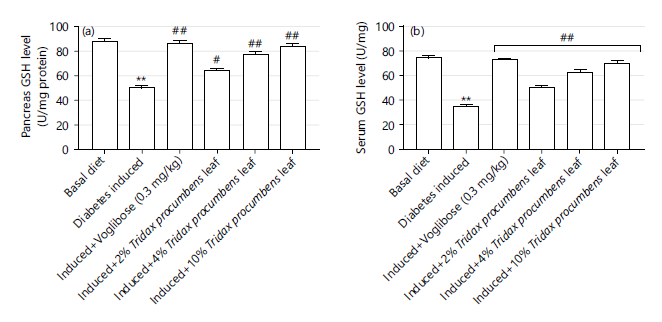
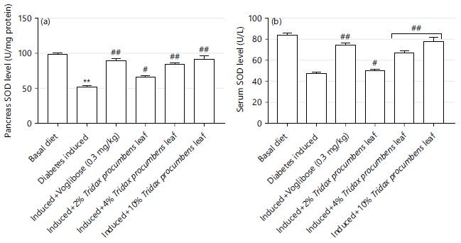
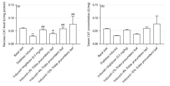
In the present study, pancreas, intestinal and serum superoxide dismutase (SOD), glutathione (GSH) and glutathione peroxidase (GPx) and catalase (CAT) activities were significantly (p<0.05) reduced in the diabetic control group. Administration of streptozotozin induced a collapse in the antioxidant defense as shown in decrease in antioxidant enzyme activities and increased MDA concentration compared to the normal control group as shown in Fig. 2-5. Several studies confirm that hyperglycemia is associated with decrease in antioxidant activities resulting from increased oxidative stress29. Oxidative stress is responsible for tissue damage and beta cell dysfunction.
Glutathione is the primary endogenous protective system which acts both as substrate in the glutathione peroxidase mediated destruction of hydroperoxides and as a nucleophilic scavenger of many compounds. The results in Fig. 2a shows a significant (p<0.05) increase in pancreatic and serum GSH activities (Fig. 2b) in groups fed with 2, 4 and 10% Tridax procumbens leaf diets compared with the diabetic control group. These results agree with the earlier findings30 which showed that the scavenging ability of serum glutathione was reduced in diabetic rats leading to β cell dysfunction.
|
|
Apart from GSH, other antioxidant enzymes with the ability to scavenge free radicals include superoxide dismutase, reduced glutathione and catalase. Superoxide dismutase catalyzes the dismutation of superoxide anion free radicals (O2-) into molecular oxygen and Hydrogen Peroxide (H2O2), and decreases the free radical responsible for damaging the cells at excessive levels31. Several natural enzymatic scavenging systems including superoxide dismutase which dismutases superoxide anion radicals to less toxic and non radical molecular oxygen and hydrogen peroxide exist.
|
On Fig. 3, Fig 3a shows a significant (p<0.05) increase in pancreas SOD activities in groups fed with 4% and 10%Tridax procumbens inclusive leaf diets was comparable to the standard drug treated while serum SOD (Fig. 3b) of the 2% Tridax procumbens leaf diet fed group showed no significant difference (p>0.05) compared with diabetic control group. The results obtained shows that the effect of Tridax procumbens leaf on SOD activity was dependent on the level of inclusion in diets and confirms the previous report of significant (p<0.05) elevation of serum superoxide dismutase activity in group treated with 400 mg/kg b.w. of methanolic extract of Euphorbia helioscopia compared to the diabetic control group with a nonsignifcant increase in the SOD activities in group treated with 200 mg/kg b.w. of same plant extract32.
Figure 4a-b displays a significant (p<0.05) increase in the pancreas and serum catalase activities in groups fed with 2, 4 and 10% Tridax procumbens leaf dietscompared with the diabetic control standard reactive oxygen species keeping the molecule at an optimal level within the cell, as it is a crucial component of the cellular signaling processes33. Catalase is an enzyme catalyzing the breakdown of hydrogen peroxide which is acrucial cellular signaling compound.
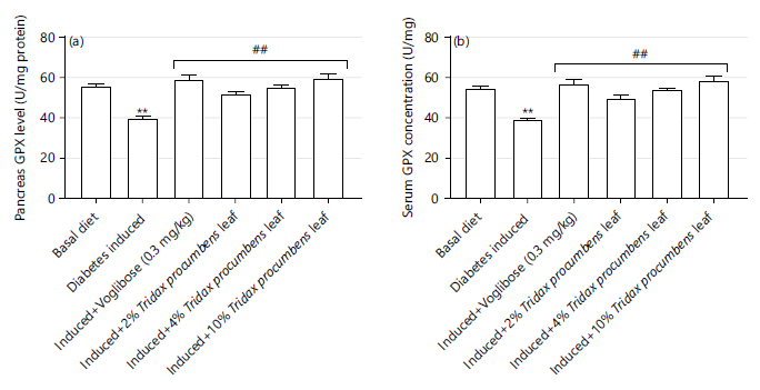
Figure 5 shows a significant (p<0.05) increase in pancreas (Fig. 5a) and serum (Fig. 5b) GPx activities in groups fed 2 and 10% Tridax procumbens leaf diets (54.07±1.01 and 58.70±1.94 U/L) as compared with diabetic control group (39.17±1.23 U/L). However, only the group fed with 10% Tridax procumbens leaf diet (58.70±1.94 U/L) showed significant elevation compared with normal control rats (54.03±1.76 U/L). Increase in serum antioxidant enzyme activities were observed as the concentration of Tridax procumbens leaf in diets increased as seen in Fig. 2-4, which shows that the administration of the diets at higher Tridax procumbens leaf inclusion levels increased the activities of serum antioxidants status markers enzymes of hyperglycemic rats.These reveal the potential of Tridax procumbens leaf to prevent the formation of free radicals in many disorders.
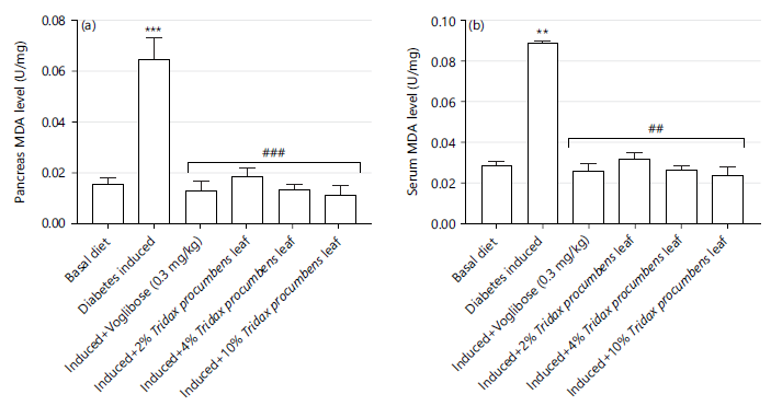
Malonaldehyde (MDA) is an aldhyde produced during the oxidative breakdown of membrane phospholipids and polyunsaturated fatty acids (PUFA)34. Lipid peroxidation assay is employed to quantify the concentration of malondialdehyde in a sample, as well as the level of MDA produced by the hydrolytic conditions of the process from lipid hydroperoxides35. As shown in Fig. 6a-b, there were significant increases in the level of MDA produced in both the pancreas and serum of the diabetic untreated group compared to the normal control group while the standard drug and Tridax procumbens leaf reduced the MDA levels in both pancreas and serum to levels comparable to that of the basal diet fed group and except for the 2% diet MDA levels were comparable to that of the standard drug group.
|
Increased MDA concentration translates to increasedfree radical generation. Development and progression in diabetes is accompanied increased oxidative stress. High free radical levels without a concomitant increase in antioxidant defense mechanism can lead to cellular damage and lipid peroxidation35. Increased hyperglycemia indced depletion in endogenous antioxidant defense system in diabetic rats may be responsible for the increased plasma and pancreatic MDA levels. Significant reduction in plasma and pancreatic MDA concentration in the Tridax procumbens leaf diet fed rats diabetic rats showed increased plasma and pancreatic MDA indicating depleted endogenous antioxidant system stemming from hyperglycemia in the Tridax procumbens leaf diet fed rats show the plant leaf as a potential lipid inhibitor via counteracting the effects of freeradicals generated.
The concentration of lipids such as Triacylglyceride (TAG), Total Cholesterol (TC), High Density Lipoprotein (HDL) Low-Density Lipoprotein (LDL), and Very Low-Density Lipoprotein (VLDL) is highly regulated to avoid certain clinical conditions such as steatosis36. This condition occurs when there is abnormal retention of lipids within a cell as a result of impairment in the normal synthesis and degradation of fats. Accumulation of these fats is often associated with disorders such as diabetes mellitus, obesity, and hepatitis C. Reduction in the level of serum HDL-cholesterol isa strong indicator of cardiovascular disease: Atherosclerosis, heart diseases Regulation of tissue lipid profile parameters is important to prevent lipid related disorders like steatosis which can result from abnormal lipid retention influencing the synthesis and degradation of fat. Fat accumulation is associated with degenerative disorders like diabetes mellitus and obesity. Lowered HDL cholesterol is a risk factor for the development of cardiovascular disorder37.
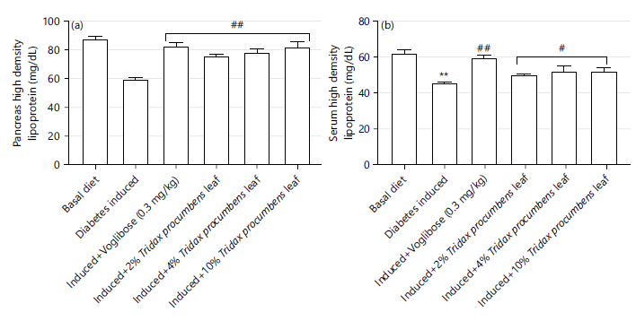
Figure 7 shows the HDL cholesterol concentration of rats fed Tridax procumbens leaf diets. The pancreatic HDL cholesterol (Fig. 7a) of rats fed Tridax procumbens diets were significantly higher than that of the untreated diabetic rats but were comparable to that of rats fed with standard drug (Voglibose 0.3 mg/kg b.w.) whereas serum HDL cholesterol of rats fed the leaf diet were significantly higher (p<0.05) than the induced untreated rats (Fig. 7b).
|
|
Rats fed 10% Tridax diet showed the highest HDL concentration compared to the diabetic untreated group. High density lipoprotein cholesterol is associated with its ability to uptake and return surplus cholesterol from peripheral tissues back to the liver and thus, to its role in prevention of atherosclerosis, transient ischemic attack, stroke and obesity38. Compared with the untreated rats, HDL cholesterol concentration was found at high concentration in rats fed 10% Tridax procumbens diets. Prevention of athreosclerosis development has been linked to the ability of HDL cholesterol to uptake and return surplus cholesterol from the peripheral tissues to the liver.
Therefore, repeated consumption of Tridax procumbens inclusive diet showed potentials in decreasing the risk of atherosclerosis development.
|
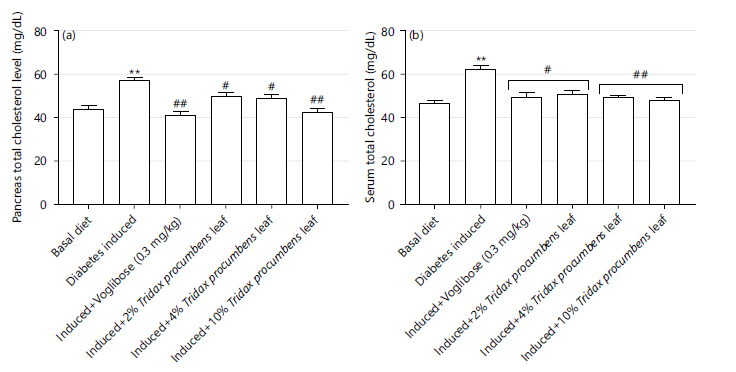
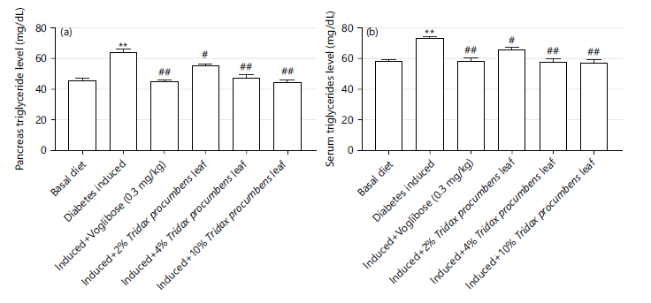
The effect of Tridax procumbens leaf diet on pancreas (Fig. 8a) and serum (Fig. 8b) total cholesterol of experimental rats. The standard drug (voglibose) group and 10% Tridax procumbens leaf diet groups showed a significant decrease (p<0.05) in total cholesterol concentration compared to pancreas and serum total cholesterol concentration in diabetic induced rats.There were no significant differences between the serum total cholesterol of the drug treated group and all the Tridax procumbens leaf inclusive dietary groups. These findings are consistent with previous reports which showed beneficial effect of Tridax procumbens extract on rat cholesterol concentration39. Figure 9(a-b) depict the triglycerides concentration distribution in pancreas and serum of experimental rats. Diabetes induction led to a significant increase (p<0.05) in triglycerides concentration in the pancreas and serum compared to the basal diet fed groups. However, the leaf diets showed significant (p<0.05) triglyceride concentration lowering effect comparable to the standard drug treated group in both tissues.
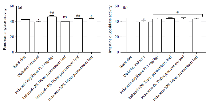
Alpha glucosidase, distributed on the brush edge of the small intestine mucosa significantly affects glycosyl structure and α-amylase are enzymes that hydrolyze (1,4)-α-D-glucosidic linkages in polysaccharides containing three or more (1,4)-α-linked D-glucose. These enzymes have the ability to hydrolyze glycosidic bonds in different sugar compounds resulting in monosaccharides, oligosaccharides, or glycosaminoglycans that raise postprandial blood glucose levels. The inhibitory effects of Tridax procumbens leaf inclusive diets on pancreatic amylase and intestinal α-glucosidase activities in induced hyperglycemic rats are shown on Fig. 10, Tridax procumbens diets significantly (p<0.05) inhibited the activities of pancreatic amylase (Fig. 10a) and intestinal glucosidase (Fig. 10b) with exception of 2% Tridax procumbens leaf diet fed rats. Hence, slowing down of carbohydrate digestion due to inhibition of α-glucosidase and amylase activities tend to lower the blood absorption of glucose and regulates blood sugar levels.This inhibition is seen as a crucial clinical validation target for the treatment of non-insulin-dependent diabetic mellitus.
Tumor Necrosis Factor-alpha (TNF-α), a pro-inflammatory cytokine produced by macrophages and monocytes, is prominently present during inflammatory processes40. It plays a crucial role in promoting bone resorption by directly activating osteoclast precursor cells via the RANKL signaling pathway41, TNF-α antagonists are used to treat moderate to severe disease in patients who cannot respond to conventional systemic therapies, have contraindications, or experience side effects.
|
|
Inflammation, a complex biological response of the immune system is indicated by increased level of Tumor Necrosis Factor-alpha (TNF-α) which plays a critical role in bone resorption. Non-responsiveness to conventional systemic therapies are usually treated with anti TNF therapy.
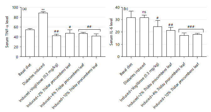
Elevated levels of TNF-α are linked to the severity of the disease including uncontrolled inflammatory responses, increased formation of osteoclast precursors, and enhanced osteoclast activity, ultimately resulting in bone resorption42. Figure 11 shows a statistically significant (p<0.05) increase in the serum levels of the pro-inflammatory cytokine TNF-α (Fig. 11a) in induced hyperglycemic untreated rats which significantly reduced in group fed 2, 4 and 10% Tridax procumbenleaf inclsive diets. The serum levels of IL-6 (Fig. 11b) in the induced hyperglycemc untreated group (31.72±2.13) were significantly decreased (p<0.05) in groups fed with 2% (21.36±2.58), 4% (17.34±1.92), and 10% (18.02±1.10) Tridax procumbens inclusive diets. Increase in the serum level of IL-6 in the induced untreated group could be due to oxidative stress which might have resulted from damage of the pancreatic cells leading to lack of insulin and hence, high concentration of circulating blood glucose43. Tridax procumbens reduced the levels of Tumor Necrosis Factor-alpha (TNF-α) and interleukin (IL)-6 in the serum of test rats compared to the diabetic untreated rat group.
CONCLUSION
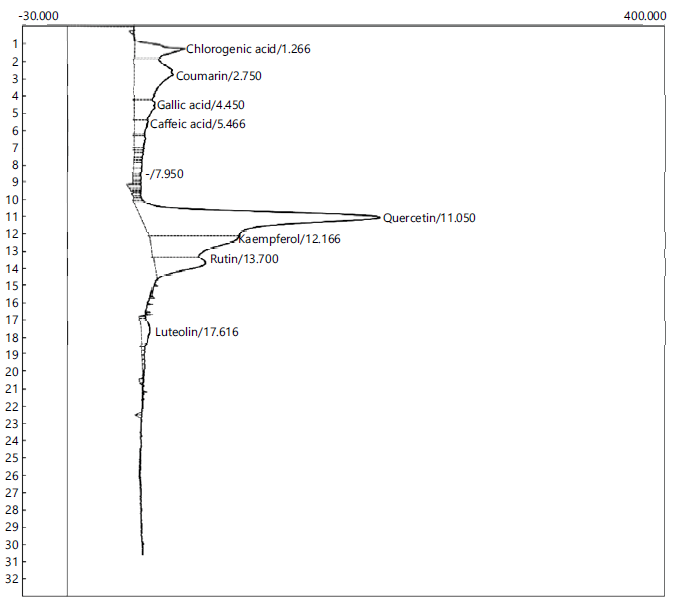
In conclusion, management of hyperglycemic rats with the Tridax procumbens leaf inclusive diets improved the antioxidant activities, lipid profile, pro-inflammatory factors and inhibited the activities of carbohydrate metabolizing enzymes in experimental rats in a concentration dependent manner. The various activities could be traced to the presence of certain bioactives such as gallic acid, qucertin, kaempferol and rutin in the plant leaf (Fig. 12). Hence, the inclusion of Tridax procumbens leaf as part of a healthy diet can be considered as a promising alternative therapeutic option and functional food for the preventoin and/or treatment of diabetic complications. This research provides a biochemical rationale for clinical investigations to further affirm these findings.
The foregoing shows the Tridax procumbens leaf contains an array of phenolic bioactives with hypoglycemic, hypolipidemic and antioxidative potentials which will find applications as phytotherapeutic alternatives to orthodox medications for the management of various degenerative disorders.
SIGNIFICANCE STATEMENT
This study explores the potential of the Tridax procumbens leaf as a source of bioactive compounds and highlights its hypoglycemic, hypolipidemic, and antioxidant activities in Streptozotocin (STZ)- induced diabetic rats. Findings from this study provide a scientific basis for the folkloric use of the plant leaf as a natural herbal neutraceutical remedy with functional properties, as opposed to synthetic drugs, for the management of hyperglycemia- and oxidative stress-related disorders.
ACKNOWLEDGMENT
The Authors thank all colleagues for their technical expertise.
REFERENCES
- Roden, M. and G.I. Shulman, 2019. The integrative biology of type 2 diabetes. Nature, 576: 51-60.
- Ahmad, E., S. Lim, R. Lamptey, D.R. Webb and M.J. Davies, 2022. Type 2 diabetes. Lancet, 400: 1803-1820.
- American Diabetes Association, 2009. Diagnosis and classification of diabetes mellitus. Diabetes Care, 32: S62-S67.
- Etxeberria, U., A.L. de la Garza, J. Campión, J.A. Martínez and F.I. Milagro, 2012. Antidiabetic effects of natural plant extracts via inhibition of carbohydrate hydrolysis enzymes with emphasis on pancreatic alpha amylase. Expert Opin. Ther. Targets, 16: 269-297.
- Ogurtsova, K., J.D. da Rocha Fernandes, Y. Huang, U. Linnenkamp and L. Guariguata et al., 2017. IDF Diabetes Atlas: Global estimates for the prevalence of diabetes for 2015 and 2040. Diabetes Res. Clin. Pract., 128: 40-50.
- Oguntibeju, O.O., 2019. Medicinal plants and their effects on diabetic wound healing. Vet. World, 12: 653-663.
- Chaudhari, H.C. and K.P. Patil, 2022. A review on medicinal importance of Tridax procumbens Linn. Res. Rev. Pharm. Pharm. Sci., 11: 1-20.
- Sofowora, A., 1993. Medicinal Plants and Traditional Medicine in Africa. 2nd Edn., Spectrum Books, Ibadan, Nigeria, ISBN-13: 9789782462190, Pages: 289.
- Parama, D., M. Boruah, K. Yachna, V. Rana and K. Banik et al., 2020. Diosgenin, a steroidal saponin, and its analogs: Effective therapies against different chronic diseases. Life Sci., 260.
- Trease, E.G. and W.C. Evans, 1978. Pharmacognosy. 11th Edn., Balliere Tindall, London, United Kingdom, ISBN: 978-0702006562, Pages: 795.
- Edeoga, H.O., D.E. Okwu and B.O. Mbaebie, 2005. Phytochemical constituents of some Nigerian medicinal plants. Afr. J. Biotechnol., 4: 685-688.
- Benderitter, M., V. Maupoil, C. Vergely, F. Dalloz, F. Briot and L. Rochette, 1998. Studies by electron paramagnetic resonance of the importance of iron in the hydroxyl scavenging properties of ascorbic acid in plasma: Effects of iron chelators. Fundam. Clin. Pharmacol., 12: 510-516.
- Ōyanagui, Y., 1984. Reevaluation of assay methods and establishment of kit for superoxide dismutase activity. Anal. Biochem., 142: 290-296.
- Sinha, A.K., 1972. Colorimetric assay of catalase. Anal. Biochem., 47: 389-394.
- Anderson, M.E., 1985. Determination of Glutathione and Glutathione Disulfide in Biological Samples. In: Glutamate, Glutamine, Glutathione and Related Compounds, Methods in Enzymology, Meister, A. (Ed.). Academic Press, New York, pp: 548-555.
- Mohandas, J., J.J. Marshall, G.G. Duggin, J.S. Horvath and D.J. Tiller, 1984. Differential distribution of glutathione and glutathione-related enzymes in rabbit kidney: Possible implications in analgesic nephropathy. Biochem. Pharmacol., 33: 1801-1807.
- Hørder, M., R.C. Elser, W. Gerhardt, M. Mathieu and E.J. Sampson, 1990. International federation of clinical chemistry: Scientific division, committee on enzymes IFCC methods for the measurement of catalytic concentration of enzymes Part 7. IFCC method for creatine kinase (ATP: Creatine N-phosphotransferase, EC 2.7.3.2). IFCC recommendation. Clin. Chim. Acta, 190: S4-S17.
- Schumann, G., R. Klauke, F. Canalias, S. Bossert-Reuther and P.F.H. Franck et al., 2011. IFCC primary reference procedures for the measurement of catalytic activity concentrations of enzymes at 37°C. Part 9: Reference procedure for the measurement of catalytic concentration of alkaline phosphatase: International Federation of Clinical Chemistry and Laboratory Medicine (IFCC) Scientific Division, Committee on Reference Systems of Enzymes (C-RSE)1. Clin. Chem. Lab. Med., 49: 1439-1446.
- Oyetayo, F.L., S.F. Akomolafe and I.A. Odeniyi, 2019. Effects of dietary supplementation of Chrysophyllum albidum fruit pulp powder on some biochemical parameters in a type 2 diabetes rat model. Vegetos, 32: 190-199.
- Bucolo, G. and H. David, 1973. Quantitative determination of serum triglycerides by the use of enzymes. Clin. Chem., 19: 476-482.
- Allain, C.C., L.S. Poon, C.S.G. Chan, W. Richmond and P.C. Fu, 1974. Enzymatic determination of total serum cholesterol. Clin. Chem., 20: 470-475.
- Lopes-Virella, M.F., P. Stone, S. Ellis and J.A. Colwell, 1977. Cholesterol determination in high-density lipoproteins separated by three different methods. Clin. Chem., 23: 882-884.
- Friedewald, W.T., R.I. Levy and D.S. Fredrickson, 1972. Estimation of the concentration of low-density lipoprotein cholesterol in plasma, without use of the preparative ultracentrifuge. Clin. Chem., 18: 499-502.
- Adeneye, A.A., 2007. Hypoglycemic and hypolipidemic effects of methanol seed extract of Citrus paradisi Macfad (Rutaceae) in alloxan-induced diabetic Wistar rats. Niger. Q. J. Hosp. Med., 18: 211-215.
- Kim, S.H., S.H. Jo, Y.I. Kwon, and J.K. Hwang, 2011. Effects of onion (Allium cepa L.) extract administration on intestinal α-glucosidases activities and spikes in postprandial blood glucose levels in SD rats model. Int. J. Mol. Sci., 12: 3757-3769.
- Kelley, W.T., D.L. Coffey and T.C. Mueller, 1994. Liquid chromatographic determination of phenolic acids in soil. J. AOAC Int., 77: 805-809.
- Thurber, M.D. and J.W. Fahey, 2009. Adoption of Moringa oleifera to combat under-nutrition viewed through the lens of the diffusion of innovations theory. Ecol. Food Nutr., 48: 212-225.
- Bobadoye, M.F., O.O. Bamisi and V.N. Enujiugha, 2016. Hypolipidemic and antioxidative effects of African star apple juice (Chrysophylum albidum) on rats fed on diets high in cholesterol and oil. Food Nutr. Sci., 7: 825-843.
- Prior, R.L., X. Wu and K. Schaich, 2005. Standardized methods for the determination of antioxidant capacity and phenolics in foods and dietary supplements. J. Agric. Food Chem., 53: 4290-4302.
- Khan, S., Hidayat Ullah Khan, F.A. Khan, A. Shah and Abdul Wadood et al., 2022. Anti-Alzheimer and antioxidant effects of Nelumbo nucifera L. alkaloids, nuciferine and norcoclaurine in alloxan-induced diabetic albino rats. Pharmaceuticals, 15.
- Bila, I., O. Dzydzan, I. Brodyak and N. Sybirna, 2019. Agmatine prevents oxidative-nitrative stress in blood leukocytes under streptozotocin-induced diabetes mellitus. Open Life Sci., 14: 299-310.
- Mustafa, I., H. Anwar, S. Irfan, H. Muzaffar and M.U. Ijaz, 2022. Attenuation of carbohydrate metabolism and lipid profile by methanolic extract of Euphorbia helioscopia and improvement of beta cell function in a type 2 diabetic rat model. BMC Complementary Med. Ther., 22.
- Nandi, A., L.J. Yan, C.K. Jana and N. Das, 2019. Role of catalase in oxidative stress- and age-associated degenerative diseases. Oxid. Med. Cell. Longevity, 2019.
- Jung, S., K.C. Nam and C. Jo, 2016. Detection of malondialdehyde in processed meat products without interference from the ingredients. Food Chem., 209: 90-94.
- Trevisan, M., R. Browne, M. Ram, P. Muti, J. Freudenheim, A.M. Carosella and D. Armstrong, 2001. Correlates of markers of oxidative status in the general population. Am. J. Epidemiol., 154: 348-356.
- Santos, A.L. and G. Preta, 2018. Lipids in the cell: Organisation regulates function. Cell. Mol. Life Sci., 75: 1909-1927.
- Fox, J.M., F. Kausar, A. Day, M. Osborne and K. Hussain et al., 2018. CXCL4/platelet factor 4 is an agonist of CCR1 and drives human monocyte migration. Sci. Rep., 8.
- Bailey, A. and S.S. Mohiuddin, 2022. Biochemistry, High Density Lipoprotein. StatPearls Publishing, Treasure Island, Florida.
- Sammeta, K., P. Kota, J. Panda, D.V.S.N. Adithya and S. Chowdary et al., 2014. Antihyper lipidemic activity of ethanolic extract of Tridax procumbens L. Int. J. Pharm. Chem. Biol. Sci., 4: 874-877.
- Saddala, M.S. and H. Huang, 2019. Identification of novel inhibitors for TNFα, TNFR1 and TNFα-TNFR1 complex using pharmacophore-based approaches. J. Transl. Med., 17.
- Zamri, F. and T.J. de Vries, 2020. Use of TNF inhibitors in rheumatoid arthritis and implications for the periodontal status: For the benefit of both? Front. Immunol., 11.
- de Vries, T.J., I. El Bakkali, T. Kamradt, G. Schett, I.D.C. Jansen and P. D'Amelio, 2019. What are the peripheral blood determinants for increased osteoclast formation in the various inflammatory diseases associated with bone loss? Front. Immunol., 10.
- Kakkar, M., T. Behl, C. Vargas-de-la Cruz, H.A. Makeen and M. Albratty et al., 2022. Tridax procumbens ameliorates streptozotocin-induced diabetic neuropathy in rats via modulating angiogenic, inflammatory, and oxidative pathways. Evidence-Based Complementary Altern. Med., 2022.
How to Cite this paper?
APA-7 Style
Oyetayo,
F.L., Odesanmi,
E.O., Akomolafe,
E.E., Afolabi,
S.A. (2026). Bioactive Constituents, Hypoglycemic, Antioxidative and Hypolipidemic Potentials of Tridax procumbens Plant Leaf Diets in Experimental Rats. Trends in Biological Sciences, 2(4), 370-385. https://doi.org/10.21124/tbs.2026.370.385
ACS Style
Oyetayo,
F.L.; Odesanmi,
E.O.; Akomolafe,
E.E.; Afolabi,
S.A. Bioactive Constituents, Hypoglycemic, Antioxidative and Hypolipidemic Potentials of Tridax procumbens Plant Leaf Diets in Experimental Rats. Trends Biol. Sci 2026, 2, 370-385. https://doi.org/10.21124/tbs.2026.370.385
AMA Style
Oyetayo
FL, Odesanmi
EO, Akomolafe
EE, Afolabi
SA. Bioactive Constituents, Hypoglycemic, Antioxidative and Hypolipidemic Potentials of Tridax procumbens Plant Leaf Diets in Experimental Rats. Trends in Biological Sciences. 2026; 2(4): 370-385. https://doi.org/10.21124/tbs.2026.370.385
Chicago/Turabian Style
Oyetayo, Folake, Lucy, Elijah Olalekan Odesanmi, Eniola Elizabeth Akomolafe, and Sheriff Ayodeji Afolabi.
2026. "Bioactive Constituents, Hypoglycemic, Antioxidative and Hypolipidemic Potentials of Tridax procumbens Plant Leaf Diets in Experimental Rats" Trends in Biological Sciences 2, no. 4: 370-385. https://doi.org/10.21124/tbs.2026.370.385

This work is licensed under a Creative Commons Attribution 4.0 International License.