
Identification of Fungal Pathogens Causing Foliar Diseases of Maize (Zea mays L.) in Makurdi Farmlands
-
Kator Liamngee


Department of Biological Sciences, Rev. Fr. Moses Orshio Adasu University, Makurdi, Benue State, Nigeria
Fayinminu Akintade OjoDepartment of Biological Sciences, Rev. Fr. Moses Orshio Adasu University, Makurdi, Benue State, Nigeria
| Received 11 Jan, 2026 |
Accepted 21 Jul, 2026 |
Published 30 Sep, 2026 |
Background and Objective: Maize production is often limited by foliar fungal diseases that damage leaves and reduce yield. Fungi causing foliar disease of maize (Zea mays L.) on farmlands in Makurdi were investigated to identify the pathogens responsible and determine their incidence and occurrence. Materials and Methods: Maize leaves showing disease symptoms were collected from three farmlands in Makurdi: Behind Modern Market (BMM), Behind Customary Court (BCC), and Terwase Agbadu Extension (TAE). Diseased leaf pieces were surface-sterilized in 5% sodium hypochlorite for 30 sec to 1 min and cultured on Potato Dextrose Agar (PDA) for fungal isolation. Pathogenicity tests were conducted on healthy maize leaves. Data collected included fungal incidence, mean fungal count, and occurrence. Statistical analyses were performed using Chi-square (χ2) and One-way Analysis of Variance (ANOVA), and means were separated using Fisher’s Least Significant Difference at 5% significance level. Results: Three fungi were isolated: Bipolaris sp., Curvularia sp., and Puccinia sorghi. Out of 275 plants sampled, 88 (32.0%) were infected. At BCC, 30/105 plants (28.8%) were infected; at BMM, 36/90 plants (39.6%); and at TAE, 22/80 plants (27.5%). There were no significant differences in fungal incidence across locations (χ2 = 3.61, df = 2; p = 0.16) or occurrence (χ2 = 1.25, df = 4, p = 0.86). Fungal occurrence was highest at BMM (37.5%), followed by TAE (33.3%) and BCC (29.2%). Curvularia sp. had the highest occurrence (41.7%), followed by Bipolaris sp. (33.3%) and P. sorghi (25%). Pathogenicity tests confirmed that these fungi caused foliar disease in maize. Conclusion: The isolated fungi were responsible for foliar disease in maize. Farmers are advised to remove residues of previous harvests before planting new crops to reduce inoculum transfer and disease incidence.
| Copyright © 2026 Liamngee and Ojo. This is an open-access article distributed under the Creative Commons Attribution License, which permits unrestricted use, distribution, and reproduction in any medium, provided the original work is properly cited. |
INTRODUCTION
Maize (Zea mays L.), which is a staple crop, that forms the foundation of daily diets and global food security, is produced and consumed by most households in West Africa1. Maize production is of utmost importance to the Nigerian economy, considering its commercial prominence and food values. It is the most cultivated crop in Nigeria in terms of land mass, with a total of 12,403,330 ha2. Africa had a total production of about 90 mL metric tonnes in 2019 and Nigeria produced about 11 million metric tonnes making it the 2nd largest producer in the continent3. In Nigeria, the major maize producing areas are Katsina, Borno, Niger, Gombe Figau, Bauchi, Kogi, Taraba, Kaduna and Oyo which accounts for 64% of maize produced in the country. The crop is suited and grows well of the ecological zones of West Africa4. As a major source of carbohydrates, it is used widely as food for humans, feed for livestock and for poultry, which has led to an increase in its cultivation in recent times5. It is a cash crop and an industrial raw material used to produce crayons, soaps, fructose syrup, dextrose, maize oil, cosmetics, biodegradable plastics, absorbent material for nappy/diaper starch, pulp abrasive, baby cereals, livestock feeds, food additives, food supplements and beverages6,7. It is also used to make organic fuel which reduces air pollution8. In Africa, an ever-growing population has become a topic of great concern with regards to food security leading to increased annual imports of maize and its products in recent times9.
Fungi are ubiquitous nature and their relationship with several hosts could either be beneficial or harmful. Fungal infection of grains before and after harvest causes reduced vigour, loss of germination, mustiness, mycotoxin contamination and moldy smell amongst others10. Many maize plantations are exposed to a variety of fungal infections. The common foliar diseases on maize plants caused by fungi include common corn rust caused by Puccinia sp., eyespot caused by Kabatiella zeae and Northern corn leaf blight caused by Exserohilum turcicum. The lesions caused by these fungi become numerous that the maize leaves are destroyed leading to yield loss11. There is a paucity of data regarding the foliar infection of maize plants on the field in Makurdi hence the need to investigate fungi causing foliar disease on maize plants on farmlands in the study area.
MATERIALS AND METHODS
Experimental location: The study was carried out at the Botany Laboratory of Biological Sciences Fr. Adasu University, Makurdi from April 2025 to October 2025. Makurdi is located on Latitudes 7'30°N and 70’45°N, Longitudes 8'30°E and 8'35°E of the equator with mean monthly temperature ranges of 27-38°C and mean annual rainfall ranges of 150-180 mm. The vegetation is guinea savannah suitable for grazing animals and farming activities which include the cultivation of crops like soybeans, maize, spinach, tobacco12.
Collection of samples: Collection of samples was done during the 2024 planting season. Leaf samples were collected from privately owned farms at three locations Behind Customary Court (BCC), Behind Modern Market (BMM) and Terwase Agbadu Extension (TAE) where maize was cultivated in Makurdi. Samples of maize leaves showing lesion, discolouration and spots were harvested. Collection of samples was done using the quadrant sampling method. In this method, the field was divided into smaller sections or blocks and ensuring each block was relatively homogenous consisting of diseased and healthy maize plants. Maize leaves with signs of fungi disease were harvested from maize plants in each quadrant on the different farmlands. The number of diseased maize plants were recorded for each quadrant. Leaves of maize showing disease symptoms were harvested in triplicates from each quadrant and brought to the laboratory. Samples collected from each quadrant were packaged separately in a polythene bags, labelled appropriately and transported to the Botany Laboratory of Rev. Fr. Moses Orshio Adasu University Makurdi for fungi isolation.
Disease incidence was assessed by expressing the number of diseased maize plants as a percentage of the total number of maize plants: These were applied in the formula13:
where, X is number of diseased plants and N is total number of plants sampled
Preparation of culture media: The Potato Dextrose Agar was prepared according to the manufacturer’s recommended procedures and used for the isolation of fungi. About 39.6 g of powdered PDA medium were dissolved in 1000 mL of sterile distilled water and stirred vigorously to homogenize. The flask content was heated on a heating mantle until the solution became clear. After heating, the flask was covered with foil paper and autoclaved at 121°C for 15 min at 760 mmHg. The sterile medium was allowed to cool to a temperature at which it could be held with hands and two to three drops of streptomycin sulphate were added to inhibit bacterial growth. The medium was poured into the Petri dishes and allowed to solidify before being used for culturing fungi.
Isolation of fungi from infected maize leaves: Fungi infecting maize leaves were isolated by cutting small sections along the margin of the diseased maize leaves. The sections were surfaced sterilized in 5% sodium hypochlorite solution for 30 sec 1 min14. Afterwards, they were rinsed in three changes of sterile distilled water to remove residuals of the sodium hypochlorite and blotted dry on sterile Whatman’s filter papers. They were then placed on solidified PDA medium. Three replicates were made for each sample. The inoculated Petri Figs were incubated at ambient temperature and observations were made daily for possible microbial growth. After 5-7 days of growth, subculturing was done to obtain pure cultures of the isolates as reported15. The data collected include.
Mean fungi count: Mean fungi count was determined by counting the number of fungi colonies that occurred in each treatment.
Occurrence of specific fungi (%): Figs were observed for growth and the occurrence of specific fungi was determined by counting the number of times each individual fungus occurred divided by the total number of fungi and expressed as a percentage using the formula16:
Subculturing of fungi isolates: To subculture, a sterilized inoculation needle was used to pick a little quantity of the fungal growth on the old culture and transferred to the centre of a freshly prepared PDA in another Petri dish. Sub-culturing was done repeatedly until pure cultures of each fungal isolate was obtained17.
Identification of fungi: Macroscopic and microscopic identification was carried out. For macroscopic identification, the colour, nature of growth, and growth rate of the fungi were observed on the PDA in Petri Figs. Microscopic identification was done by staining a glass slide with a drop of Lactophenol in cotton blue and with the aid of an inoculation needle a small quantity of the fungal colony was placed on the stained-glass slide, covered with a cover slip and viewed under the 40× objective lens of the light microscope. The observed characteristics of the fungi were compared with a standard chart for identification18.
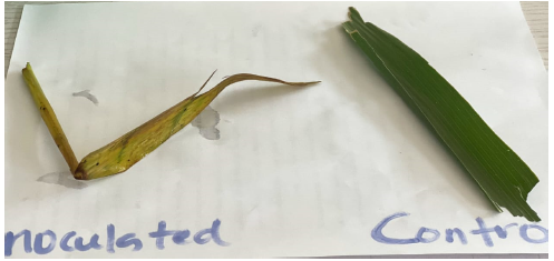
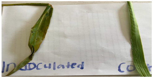
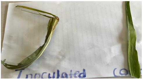
Pathogenicity of fungal isolates on healthy maize leaves in Makurdi: Healthy maize leaves were washed with sterile distilled water and thereafter sterilized in 5% sodium hypochlorite solution for 30 sec. Mycelia discs of fungal isolates from a 2 week old culture were used to inoculate the maize leaves. All the leaves were inoculated except the control. To inoculate the leaves, the surface of the pure culture was flooded with 5 mls of sterile distilled water and allowed to settle. The suspension was poured into a clean bowl and each leaf sample was immersed and allowed to stay for 30 sec–1 min. After inoculation, the leaves were placed into a disposable nylon containing little quantity of sterile distilled water to maintain humidity. Thereafter, it was placed inside the trays and held tight using a rubber band. Controls were maize leaves immersed in sterile distilled water only. After 5-7 days of post inoculation, physical examination of the leaves was used to ascertain the symptoms of disease. On appearance of symptoms, the tissues at the margin of the healthy and diseased parts were excised, sterilized and placed on PDA and incubated at room temperature for 5-7 days. At the end of this period, morphological characteristics and growth patterns observed in each case were compared with the ones of the original isolates.
Data analysis: Data obtained from the study were analyzed using Chi-square and Analysis of Variance. Means were separated using Fisher’s Least Significant Difference at 5% level of significance.
RESULTS
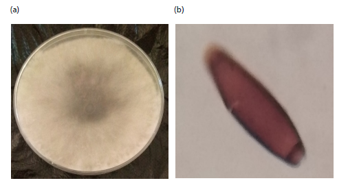
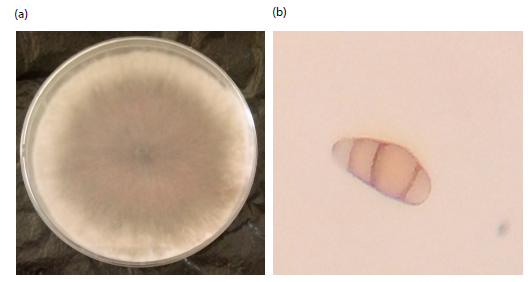
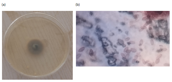
The fungi isolated causing foliar disease of maize plants on farmlands in Makurdi were identified as; Curvularia sp., Bipolaris sp. and Puccinia sorghi. The macroscopic and microscopic views of the organisms are presented in Fig. 1-3(a-b). The colony of Bipolaris sp., on PDA was initially white later turning to dark brown with cottony like appearance (Fig. 1a). Microscopically, the conidia of Bipolaris sp., were brown in colour, elongated and cylindrical (Fig. 1b). The colony of Curvularia sp., were velvety in appearance with a dark centre (Fig. 2a). When viewed under the microscope, the conidia were brown in colour, curved and septate (Fig. 2b). Puccinia sorghi colony on PDA was darkish green in colour (Fig. 3a). The conidia were ovoid and transparent and formed clusters (Fig. 3b).
Incidence of fungi causing foliar disease on maize plants on farmlands in Makurdi: The incidence of fungi causing foliar disease on maize plants in farmlands within Makurdi is presented in Table 1. Results obtained showed that out of the two hundred and seventy-five plants sampled, 88 (32.00%) were infected with fungi. Behind Customary Court (BCC), 30 (28.80%) of the 105 plants had diseased leaves, while 36 (39.60%) out of the 90 plants Behind Modern Market (BMM) had infected leaves. A total of 22 (27.50%) plants out of the 80 plants sampled at Terwase Agbadu Extension (TAE) had diseased leaves. There was no significant relationship in the incidence of fungi on diseased maize leaves across the locations (χ2 = 3.61, df = 2, p = 0.16).
Mean fungi count on maize leaves collected from farmlands in Makurdi: The mean fungi count on maize leaves from farmlands in Makurdi is shown in Table 2. Results obtained showed higher fungi count on maize leaves collected at BMM (3.00) compared to TAE (2.67) and BCC (2.33). There was no significant difference in mean fungi count on maize leaves between the locations.
Occurrence of specific fungi on maize leaves on farmlands: The occurrence of specific fungi on maize leaves on farmlands within Makurdi is shown in Table 3. Curvularia sp., had the highest occurrence 4 (44.4%) on maize leaves in Behind Modern Market (BMM), followed by Bipolaris sp., 3 (33.3%) and P. sorghi 2 (22.2%). At Behind Customary Court (BCC), Curvularia sp. and Bipolaris sp., were the dominant fungi each with 3, accounting for 42.90%, followed by P. sorghi 1(14.30%). In Terwase Agbadu Extension (TAE), Curvularia sp., and P. sorghi had the highest occurrence each with 3 (37.50%) and Bipolaris sp., 2 (25.00%) being the least occurrence. Overall, Curvularia sp., had higher occurrence 10 (41.70%), followed by Bipolaris sp., 8 (33.30%) and P. sorghi 6 (25.00%), respectively. Maize eaves at Behind Modern Market recorded the highest occurrence of fungi 9 (37.50%) followed by Terwase Agbadu Extension 8 (33.30%) and Behind Customary Court 7 (29.20%) respectively.
|
|
|
| Table 1: | Incidence of fungi causing foliar disease on maize plants on farmlands in Makurdi | |||
| Location | Number examined | Number infected (%) |
| BMM | 90 | 36 (39.6) |
| BCC | 105 | 30 (28.8) |
| TAE | 80 | 22 (27.5) |
| Total | 275 | 88 (32.0) |
| χ2 = 3.61, df = 2, p = 0.16, BMM: Behind Modern Market, BCC: Behind Customary Court and TAE: Terwase agbadu extension | ||
| Table 2: | Mean fungi count on maize leaves collected from farmlands in Makurdi | |||
| Location | Fungi count |
| BMM | 3 |
| BCC | 2.33 |
| TAE | 2.67 |
| FLSD (0.05) | NS |
| FLSD: Fisher’s Least Significant Difference; NS: Not Significant, BMM: Behind modern market, BCC: Behind customary court and TAE: Terwase agbadu extension | |
|
|
|
| Table 3: | Occurrence of specific fungi on maize leaves based on farmlands | |||
| Fungi dpecies | ||||
| Location | Curvularia sp., | Bipolaris sp., | P. sorghi | Total (%) |
| BMM | 4 (44.4) | 3 (33.3) | 2 (22.2) | 9 (37.5) |
| BCC | 3 (42.9) | 3 (42.9) | 1 (14.3) | 7 (29.2) |
| TAE | 3 (37.5) | 2 (25.0) | 3 (37.5) | 8 (33.3) |
| Total | 10 (41.7) | 8 (33.3) | 6 (25.0) | 24 (100.0) |
| χ2 = 1.25, df = 4, p = 0.86, BMM: Behind modern market, BCC: Behind customary court and TAE: Terwase agbadu extension | ||||
| Table 4: | Morphological description of disease symptoms produced by fungal isolates on healthy maize leaves | |||
| Fungal organisms | Symptoms produced |
| Curvularia sp., | Leaves produced small, tan coloured lesions with brown margins that were surrounded by ayellowish halo (Fig. 4) |
| Bipolaris sp., | Lesions produced brown colouration that elongate between leaf veins (Fig. 5) |
| Puccinia sorghi | Leaves became small, yellow and twisted (Fig. 6) |
Chi-square analysis showed that there was no statistically significant relationship in occurrence of fungi on maize leaves based on location χ2 = 1.25, df = 4, p = 0.86 shown in Table 4.
DISCUSSION
Fungi causing foliar disease of maize plants on farmlands in Makurdi in this study were; Curvularia sp., Bipolaris sp. and Puccinia sorghi. This disagrees with the findings of Bwakat et al.19 in Jos, Nigeria in which they reported Aspergillus niger, Aspergillus flavus, Fusarium sp. and Penicillium sp as fungi causing foliar disease on maize plants. However, the fungi isolated in this study is similar to the fungi reported20 in India in which they isolated Bipolaris maydis, Curvularia, Drechslera and Alternaria sp., as pathogens causing maize leaf blight (MLB). Several studies have focused on the isolation of seed borne fungi of maize and reported various fungi species21. In Kebbi State, Nigeria isolated Aspergillus niger, Mucor sp., Aspergillus fumigatus, Aspergillus terreus and Penicillium in Maize;22 isolated Aspergillus fumigatus, Aspergillus terreus, Penicillium sp., Fusarium sp. and Aspergillus niger when they studied the incidence of fungal flora and aflatoxin content of millet and maize cereal grains sold in Guinea Savanna zones of Kebbi State23. In Ethiopia isolated Aspergillus niger, Aspergillus flavus, Penicillium sp., in Maize samples;24 isolated Aspergillus niger and Aspergillus flavus from Maize seeds collected from Konshisha LGA of Benue State25. In Yola, Nigeria isolated Aspergillus ustus, Fusarium solani, Aspergillus flavus, Rhizopus stolonifer and Botrytis cinerea in Maize seeds. All the reported works on seedborne fungi have not implicated any of the fungi isolated on foliar disease of maize in this study. This suggests that Curvularia sp., Bipolaris sp. and Puccinia sorghi are peculiar in causing foliar disease of maize. The variations in the fungi reported on foliar disease of maize in this present study compared with other studies may be due to geographical locations, the environmental conditions, the soil type and the method of cropping system23.
Maize plants surveyed for foliar disease at Behind Modern Market (BMM) in this study showed the highest occurrence of fungi compared to plants surveyed in other locations. This may be due to a more favorable climate and essential nutrient for foliar disease development at BMM. These conditions can promote the growth and spread of fungal pathogens26. Another reason may be due to poor agronomic practice such as incomplete removal of inocula of previous harvest from the farmland before the new cropping season. Study by Dixon and Tilston27 stated that poor soil drainage, nutrient deficiencies, soil-borne pathogens and crop management practices can all contribute to foliar disease.
Curvularia sp., was the most frequently isolated fungi from diseased maize leaves compared to other fungi organisms as observed in this study. This may be due to the susceptible nature of the host plant to Curvularia sp. Another study by Raja et al.28 stated that Curvularia species are known to infect a wide range of plant species, including maize. Also, it was mentioned in the study of Enyiukwu et al.29 that different plant species and cultivars can vary in their susceptibility to foliar disease. The soil quality in the study location may have been conducive to Curvularia growth. Curvularia species are known to thrive in soils with high organic matter content and poor drainage27. Also, BMM may have had a low level of fungal competition, allowing Curvularia to dominate the fungal community.
The fungi isolated in this study were able to cause disease conditions when inoculated into healthy maize leaves producing different symptoms as revealed by the pathogenicity. Curvularia is a genus of fungi that includes several species that are pathogenic to plants. Curvularia sp., has been reported to cause leaf spot and blight diseases in maize and other crops28. Bipolaris is a genus of fungi that includes several species that are pathogenic to plants. Bipolaris sp., has been reported to cause leaf spot and blight diseases in maize and other crops30. Puccinia sorghi is a fungus that is commonly associated with tropical plants. It has been reported to cause dieback and decay of plant tissues, including leaves and stems31.
SIGNIFICANCE STATEMENT
This study is significant as it identifies key fungal pathogens responsible for foliar diseases of maize in Makurdi, providing essential baseline information for disease diagnosis and management. The findings will support farmers, agronomists, and extension services in developing timely and effective control strategies to reduce yield losses and improve maize productivity. In addition, the study contributes to the existing body of knowledge on maize phytopathology and regional fungal diversity, which is important for future research and integrated disease management programs.
CONCLUSION
Curvularia sp., Bipolaris sp. and Puccinia sorghi were fungi causing foliar disease of maize plants in different farmlands in Makurdi. The incidence and occurrence of fungi causing foliar disease was highest on maize leaves collected Behind Modern Market in Makurdi. Curvularia sp was the most frequently observed fungi causing foliar disease in maize. The fungi were able to induce disease symptoms when inoculated on healthy maize leaves.
Based on the findings of this study, it is recommended that farmers should regularly inspect their crops for any signs of disease and take immediate action upon detecting symptoms to prevent further spread. In addition, proper field sanitation practices should be strictly followed by thoroughly removing and clearing all remnants of the previous harvest before the onset of the new planting season, as this helps eliminate potential sources of inoculum that may infect new crops.
REFERENCES
- Amegnaglo, C.J., 2018. Determinants of maize farmers' performance in Benin, West Africa. Kasetsart J. Social Sci., 41: 296-302.
- Onyibe, J.E., B.M. Sani, D. Baba, H. Chindo, I.K. Ibrahim and M. Malumfashi, 2014. Maize: Production, Marketing, Processing & Utilization in Nigeria. National Agricultural Extension Research Liaison Services, Kaduna State, Nigeria, Pages: 39.
- Makama, S.A., S.M. Umar, M.A. Isah, M.S. Sadiq and B.D. Magaji, 2023. Socioeconomic factors influencing maize production in Giwa local government area of Kaduna State, Nigeria. J. Agric. Environ., 18: 135-139.
- Obour, P.B., I.K. Arthur and K. Owusu, 2022. The 2020 maize production failure in Ghana: A case study of Ejura-Sekyedumase Municipality. Sustainability, 14.
- Traore, I.Y., O.O. Aminu and A.E. Adekoya, 2018. Factors influencing maize production in Sikasso Region of Mali. J. Agric. Ext., 22: 31-39.
- Acharya, V. and B.R. Young, 2008. A review of the potential of bio-ethanol in New Zealand. Bull. Sci., Technol. Soc., 28: 143-148.
- Tongjura, J.D.C., G.A. Amuga and H.B. Mafuyai, 2010. Laboratory assessment of the susceptability of some varieties of Zea mays infested with Sitophilus zeamais, Motsch. (Coleoptera, Curculionidae) in Jos, Plateau State, Nigeria. Sci. World J., 5: 55-57.
- Vermerris, W., A. Saballos, G. Ejeta, N.S. Mosier, M.R. Ladisch and N.C. Carpita, 2007. Molecular breeding to enhance ethanol production from corn and sorghum stover. Crop Sci., 47: S-142-S-153.
- Mugo, S., H. de Groote, D. Bergvinson, M. Mulaa, J. Songa and S. Gichuki, 2005. Developing Bt maize for resource-poor farmers-recent advances in the IRMA project. Afr. J. Biotechnol., 3: 1490-1504.
- Kortei, N.K., A.A. Agyekum, F. Akuamoa, V.K. Baffour and H.W. Alidu, 2019. Risk assessment and exposure to levels of naturally occurring aflatoxins in some packaged cereals and cereal based foods consumed in Accra, Ghana. Toxicol. Rep., 6: 34-41.
- Ngea, G.L.N., X. Qian, Q. Yang, S. Dhanasekaran and G. Ianiri et al., 2021. Securing fruit production: Opportunities from the elucidation of the molecular mechanisms of postharvest fungal infections. Compr. Rev. Food Sci. Food Saf., 20: 2508-2533.
- Onuche, P., A. Victoria and I.A. Michael, 2023. Daily air temperature variation in Makurdi metropolis using analysis of variance model. Int. J. Sci. Res. Arch., 9: 191-200.
- Wariyo, A., M. Dejene, E. Derso and Z. Bekeko, 2025. The intensity of coffee berry disease (Colletotrichum kahawae Waller and Bridge) and its association with biophysical factors in Southern Ethiopia. J. Plant Pathol., 107: 191-202.
- Ekhuemelo, C. and E.O. Otor, 2021. Assessment of fungi associated with the leaf spot disease of fluted pumpkin (Telfairia occidentalis) Hook F.) and their management using botanicals. Niger. J. Mycol., 13: 31-43.
- Fayinminu, A.O., K. Liamngee, T. Gbira, N.G. Fru and Y. Rekiya et al., 2025. Identification and pathogenicity of fungi causing postharvest decay of Irish potato (Solanum tuberosum L.) tubers sold in Makurdi. Eur. J. Ecol. Biol. Agric., 2: 3-13.
- Kator, L., A.T. Ogo-Oluwa and A.B. Kemi, 2016. Isolation and identification of seed borne fungi of common bean (Phaseolus vulgaris L.) from selected markets in Makurdi. Int. J. Appl. Agric. Sci., 2: 75-78.
- Kator, L., F.A. Ojo, A.B. Ocheidudu, A.H. Ngodoo and Y.J. Doowuese et al., 2025. Effect of Trichoderma species on seed borne fungi of pearl millet (Pennisetum glaucum L.R. Br) using dual culture technique. Res. J. Bot., 20: 55-65.
- Barnett, H.L. and B.B. Hunter, 1972. Illustrated Genera of Imperfect Fungi. 3rd Edn., Burgess Publishing Company, Minneapolis, Minnesota, ISBN: 9780808702665, Pages: 241.
- Bwakat, E.P., P.O. Nwadiaro and I.A. Nnebechukwu, 2023. Occurrence and distribution of fungal species associated with maize in selected farms in Plateau State, Nigeria. Direct Res. J. Agric. Food Sci., 11: 206-212.
- Singh, V., D.K. Lakshman, D.P. Roberts, A. Ismaiel, A. Abhishek, S. Kumar and K.S. Hooda, 2021. Fungal species causing maize leaf blight in different agro-ecologies in India. Pathogens, 10.
- Aminu, M. and J.N. Keta, 2024. Study of fungi on stored maize (Zea mays L.) in Kebbi State, Nigeria. J. Curr. Opin. Crop Sci., 2: 55-59.
- Ma, Z., C. Liang, H. Wang, J. Liu, X. Zhou and W. Zhou, 2025. Biotic and abiotic factors influencing maize plant height. Int. J. Mol. Sci., 26.
- Tsedaley, B. and G. Adugna, 2016. Detection of fungi infecting maize (Zea mays L.) seeds in different storages around Jimma, Southwestern Ethiopia. J. Plant Pathol. Microbiol., 7.
- Fayinminu, A.O, T.T. Ati, B.L. Agbatar, S.J. San and J.J. Orbunde, 2020. Incidence of Aspergillus species in maize seeds in Konshisha Local Government of Benue State and their control using neem leaf extract. Int. J. Res. Publ., 53.
- Usman, T. and Z.G. Bawa, 2018. Identification and control of seed born fungal parhogens of maize seed (Zea mays) using plant extracts. Int. J. Sci. Res., 7: 880-883.
- Al-Masoodi, I.H., A.F.M. Al-Rubaye and H.J. Hussein, 2023. Isolation and diagnosis of the fungi associated with maize seeds collected from local markets in Karbala, Iraq. Caspian J. Environ. Sci., 21: 665-672.
- Dixon, G.R. and E.L. Tilston, 2010. Soil-Borne Pathogens and Their Interactions with the Soil Environment. In: Soil Microbiology and Sustainable Crop Production, Dixon, G.R. and E.L. Tilston (Eds.), Springer, Netherlands, ISBN: 978-90-481-9479-7, pp: 197-271.
- Raja, H.A., A.N. Miller, C.J. Pearce and N.H. Oberlies, 2017. Fungal identification using molecular tools: A primer for the natural products research community. J. Nat. Prod., 80: 756-770.
- Enyiukwu, D.N., C.C. Ononuju and J.O. Maranzu, 2018. Plant pathogenic fungi-novel agents of human diseases: Implications for public health. Greener J. Epidemiol. Public Health, 6: 001-019.
- Sivanesan, A., 1996. Corynesporasca caryotae gen. et sp. nov. with a Corynespora anamorph, and the family Corynesporascaceae. Mycol. Res., 100: 783-788.
- Zúñiga-Silvestre, C.A., C. de-León-García-de-Alba, V. Ayala-Escobar and V.A. González-Hernández, 2020. Induced resistance to common rust (Puccinia sorghi), in maize (Zea mays). Emir. J. Food Agric., 32: 11-18.
How to Cite this paper?
APA-7 Style
Liamngee,
K., Ojo,
F.A. (2026). Identification of Fungal Pathogens Causing Foliar Diseases of Maize (Zea mays L.) in Makurdi Farmlands. Trends in Biological Sciences, 2(3), 314-323. https://doi.org/10.21124/tbs.2026.314.323
ACS Style
Liamngee,
K.; Ojo,
F.A. Identification of Fungal Pathogens Causing Foliar Diseases of Maize (Zea mays L.) in Makurdi Farmlands. Trends Biol. Sci 2026, 2, 314-323. https://doi.org/10.21124/tbs.2026.314.323
AMA Style
Liamngee
K, Ojo
FA. Identification of Fungal Pathogens Causing Foliar Diseases of Maize (Zea mays L.) in Makurdi Farmlands. Trends in Biological Sciences. 2026; 2(3): 314-323. https://doi.org/10.21124/tbs.2026.314.323
Chicago/Turabian Style
Liamngee, Kator, and Fayinminu Akintade Ojo.
2026. "Identification of Fungal Pathogens Causing Foliar Diseases of Maize (Zea mays L.) in Makurdi Farmlands" Trends in Biological Sciences 2, no. 3: 314-323. https://doi.org/10.21124/tbs.2026.314.323

This work is licensed under a Creative Commons Attribution 4.0 International License.